Journal of
eISSN: 2379-6359


Case Report Volume 2 Issue 5
Department of Neurosurgery, Clinique chirurgicale de Bel Air, France
Correspondence: Keyvan Mostofi, Department of Neurosurgery, Clinique chirurgicale de Bel Air, Angouleme, France, Tel 0680575180
Received: April 05, 2015 | Published: May 28, 2015
Citation: Mostofi K. Skull and soft tissue metastasis of an occult follicular thyroid carcinoma: a case report. J Otolaryngol ENT Res. 2015;2(5):162-164. DOI: 10.15406/joentr.2015.02.00037
Follicular thyroid carcinoma (FTC) is the second most common cancer of the thyroid after papillary carcinoma. Skull metastasis of thyroid carcinoma is rare and only about 1.7% of patients with differentiated thyroid carcinoma present bone metastasis. We present a very rare case of skull and soft tissue metastasis from an occult FTC in a 60-year-old female with 4years follow-up.
Keywords: skull metastasis, follicular thyroid carcinoma, tumor
Follicular thyroid carcinoma (FTC) is the second most common cancer of the thyroid after papillary carcinoma. The incidence of FTC has been estimated to be 33-35% by recent studies.1‒7 According to Ito et al.,8 there are two types of FTC: minimally invasive FTC and widely invasive. Among them, minimally invasive FTC is most common whereas widely invasive one, which is less frequent type, is very aggressive and has a poor prognosis. FTC is more common in women and occurs in middle aged adults.6,7,9 Bone metastasis is the second frequent type of metastasis after lung metastasis.1‒7,9‒13 Skull metastasis of thyroid carcinoma is rare and only about 1.7% of patients with differentiated thyroid carcinoma present bone metastasis.3‒6 Few cases are reported. Metastasis of FTC is often osteolytic. The differential diagnosis of lytic skull lesions is skull metastasis from lung, breast and prostate.
We present a case of skull and soft tissue metastasis from an occult FTC with 4years follow-up.
A 60-year-old female patient native of St. Lucia was admitted in the department of Neurosurgery with a mass in the left parietal bone. There was no other medical history of note for this patient. The patient had discovered the mass herself three months ago. She had no other complaints. The mass measured 4cm x 5cm x 5cm approximately and was firm in consistency but lessstiff than bone tissue. Systemic and neurological examinations revealed no other abnormalities. Routine blood tests turned out to be normal.
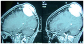
MRI demonstrated a lytic extra dural mass with contrast enhancement, eroding bone and slight cerebral tissue shift (Figure 1). Thoracoabdominal CT was normal.

Figure 1 Sagittal T1 weighted magnetic resonance image (MRI) showing the mass with invasion of the scalp (arrows).
During surgery, we found an extra dural bone lytic lesion. The dura- mater was intact and was not invaded by the tumor. On the other hand, we found tumor invasion in the scalp. In fact, the normal tissue was not recognizable beside the tumor. We removed the tumour completely and scratched and coagulated the inner layer of scalp about 2cm around the resection. Therefore the resection was macroscopically complete.
The frozen section pieces taken from the inner layer of the scalp as well as the tumour itself showed the existence of thyroid tumour cells with signs of malignancy. We therefore decided to excision of the scalp on a broad base of it (8x8cm) (Figure 2).
We performed a scalp expansion to cover the resected portion of the scalp.
Histological examination showed the metastatic origin from a follicular carcinoma of the thyroid with extracapsular spread, nuclear polymorphism and increased nucleo-cytoplasmic ratio (Figure 3).
The patient had a second Thoracoabdominal and pelvic CT which showed no metastasis. The tumour markers showed the following results: thyroglobuline 13ng/l, antigen CEA: 2,2ng/ml. Thyroid assessment demonstrated this results: TSH: 0.7 microunity/ml, calcitonine 4 pg / mL, T4: 5.5mcg/dL T3; 3 nmol/L. Calcemia was 97mg/l.
Whole-body bone scintigraphy showed no other bone metastasis. The patient also underwent a thyroid scintigraphy with iodine-123 and technetium-99 and Ultrasound-Guided Thyroid Biopsy followed by total thyroidectomy. Surgical sampling results confirmed that there was a follicular thyroid carcinoma. The patient has been followed since her surgery. She has a substitution treatment and has an annual Whole-body bone scintigraphy and Thoracoabdominal and pelvic CT followed by biannual blood tests until four years. After four years follow-up, she is fine and has no signs of metastasis elsewhere.
The rate of skull metastasis is around 15 - 25% in cancer patients.6 Tumors most frequently encountered are breast cancer, lung cancer, prostate cancer, thyroid cancer and melanoma.14 Follicular thyroid carcinoma (FTC) is the second most common cancer of the thyroid. Cranial metastasis of follicular thyroid carcinoma occurs about 1% of cases.1‒7,9,13,15 Nagamine et al.,16 reported 12 cases of skull metastasis of thyroid carcinoma and found a mean age of 60 years. There are very rare case reports of skull metastasis of thyroid follicular carcinoma 15,17‒20,21 while the incidence of Skull metastasis of occult follicular thyroid carcinoma is extremely rare.19,20,22 They are usually single, and asymptomatic. Diagnosis of the tumor is easy. The patient usually finds himself the tumor which is a palpable masse, loosely attached with firm consistency. On MRI preoperatively, it is an epidural lytic and vascular mass with irregular edge. The differential diagnosis of an osteolytic skull lesion are primary tumour like cholesteatom, epidermoid cyst, fibrous dysplasia and metastatic breast, lung, kidney cancer and neuroblastoma.22 Although the primary tumor is well differentiated, the prognosis is not good and the survival rate after 10years is around 27%.23‒26
The surgery is obviously principal treatment for solitary skull metastasis with complete excision because there is risk of recurrence in case of incomplete resection. If the dura-mater is not invaded, meticulous cleaning and coagulating is enough. If the dura is infiltrated, removal and reconstruction is necessary. A sufficient circumferential margin of scalp is recommended. Search of the original tumor and distant metastasis in particular bone metastasis is essential. There is no established consensus protocol about postoperative follow-up. In that case we recommend a regular postoperative follow up.
None.
Author declares there are no conflicts of interest.
None.

©2015 Mostofi. This is an open access article distributed under the terms of the, which permits unrestricted use, distribution, and build upon your work non-commercially.