eISSN: 2377-4304


Research Article Volume 17 Issue 5
University Women’s Hospital, Paracelsus Medical University, Nuremberg, Germany
Correspondence: Andrea Lai, University Women´s Hospital, Paracelsus Medical University, Nuremberg, Germany
Received: August 26, 2026 | Published: September 25, 2026
Citation: Lai A, Panou K, Kalfoutzos K, et al. Umbilical eversion with direct trocar insertion: a standardized transumbilical entry technique for robotic gynecologic surgery. Obstet Gynecol Int J. 2026;17(7):205-210. DOI: 10.15406/ogij.2026.17.00847
Background: Safe and efficient abdominal entry is an essential step in minimally invasive gynecologic surgery. Several entry techniques have been described, but no single approach has been universally established as superior. We describe a standardized transumbilical entry technique combining complete umbilical eversion with direct trocar insertion (DTI) for robotic-assisted gynecologic surgery.
Methods: A retrospective, single-center observational cohort study was conducted at the University Department of Gynecology, Nuremberg General Hospital, Germany, including patients who underwent robotic-assisted gynecologic surgery between January 2021 and December 2024. The technique was applied in all 401 patients included in the analysis. The umbilicus was completely everted, a median sagittal incision of approximately 1 cm was created, and an 8-mm optical trocar was directly introduced through the preformed opening along a vertical trajectory of approximately 90°. Entry time was measured by the surgical team using a stopwatch, from the beginning of the skin incision to complete insertion of the trocar into the abdominal cavity. A historical cohort of 200 patients undergoing conventional laparoscopy with Veress needle entry during the same period was used solely for descriptive comparison of entry time.
Results: The technique was successfully completed in all 401 patients, with no documented entry failures or need for conversion to an alternative access technique. Mean entry time was 45 seconds, with an observed range of 40–65 seconds. In the historical Veress cohort, entry time was 3 to < 4 minutes in 30% of patients, 4 to <5 minutes in 60%, and ≥5 minutes in 10%. No clinically relevant access-related complication was documented in the direct entry cohort. During the available postoperative follow-up, no umbilical wound infection, wound dehiscence, clinically relevant umbilical pain, or umbilical hernia related to the access site was documented. Long-term systematic surveillance for incisional or umbilical hernia was not performed.
Conclusion: Umbilical eversion combined with direct trocar insertion was feasible and reproducible within this experienced robotic gynecologic surgery team. The technique allowed rapid establishment of abdominal access and was applied in patients with varying BMI with or without previous abdominal surgery. However, the retrospective single-center design and the use of a historical comparator with different surgical indications limit conclusions regarding comparative safety or efficacy. Prospective comparative studies are warranted to further evaluate this approach.
Keywords: Robotic surgery, gynecologic surgery, laparoscopic entry, direct trocar insertion, transumbilical entry, umbilical eversion, abdominal access
Within minimally invasive surgery, a heterogeneous variety of laparoscopic entry techniques has been described. Although the incidence of serious entry-related complications is generally low, the optimal entry method and anatomical site for laparoscopic access remain subjects of ongoing discussion.1–3
The two principal methods for peritoneal access are the closed and open (Hasson) techniques, both of which are commonly performed through the umbilical region. The closed technique generally involves insertion of a Veress needle followed by pneumoperitoneum and trocar placement, whereas the open approach, originally described by Hasson, permits access to the peritoneal cavity under direct visualization.4 Alternative approaches include direct trocar entry, optical trocars, and non-umbilical access, most commonly through Palmer's point.5,6
In this context, we describe a standardized transumbilical entry technique that combines complete umbilical eversion with direct trocar insertion (DTI). The technique has been routinely used in robotic-assisted gynecologic procedures at our institution since 2021.
Previous techniques have incorporated umbilical eversion and longitudinal incision of the umbilical base. For example, Sakamoto et al. described a modified closed-entry technique in which the umbilicus was everted and its base incised longitudinally; however, their technique subsequently used a Veress needle through a Step trocar, followed by pneumoperitoneum and trocar placement.7 The technique described in the present study differs in that the fascial and peritoneal opening is created directly with closed scissors, followed immediately by insertion of the optical trocar before establishment of pneumoperitoneum.
To our knowledge, the specific standardized sequence described here - complete umbilical eversion, median sagittal skin incision, direct opening of the fascia and peritoneum with closed scissors, followed by immediate insertion of a 8-mm optical trocar before pneumoperitoneum - has not previously been described in robotic-assisted gynecologic surgery.
The technique was developed with particular consideration of patients in whom the umbilical region may be difficult to access, including patients with obesity. However, it has been routinely applied across a broad range of patient anatomies and was not restricted to a specific BMI category.
Study design and patient population
This was a retrospective, single-center observational cohort study conducted at the University Department of Gynecology, Nuremberg General Hospital, Germany. The study period extended from January 2021 to December 2024.
All patients who underwent robotic-assisted gynecologic surgery during the study period in whom the described transumbilical entry technique was used and for whom the relevant postoperative clinical documentation was available at our institution were considered for inclusion. A total of 401 patients fulfilled the study criteria and were included in the final analysis.
The cohort therefore represents all standard robotic-assisted procedures performed during the study period where the described technique was used and the relevant perioperative and postoperative documentation was available. All patients who met these predefined criteria were included in the final analysis.
The study was based on retrospectively available institutional clinical documentation. No intervention or modification of patient care was undertaken for the purposes of the study.
Data protection and confidentiality
Clinical data were retrospectively extracted from the institutional electronic medical records. The data used for the present analysis were anonymized. No patient names, surnames, or other identifying information were included in the study database or analytical tables. The data were used exclusively in anonymized form for the purposes of the retrospective analysis.
Ethics statement
This retrospective study was conducted using anonymized clinical data extracted from the institutional electronic medical records. According to the institutional assessment, approval by the ethics committee was not required for this retrospective analysis because the study used anonymized data and did not involve any intervention or modification of patient care.
Patient characteristics
The patients ranged in age from 35 to 85 years.
The age distribution was as follows: 24 patients (6.0%) were aged 35–44 years, 56 (14.0%) were aged 45–54 years, 132 (32.9%) were aged 55–64 years, 140 (34.9%) were aged 65–74 years, and 49 (12.2%) were aged 75–85 years.
BMI distribution was as follows: 148 patients (36.9%) had a BMI <25 kg/m², 156 (38.9%) had a BMI of 25–<30 kg/m², 64 (16.0%) had a BMI of 30–<35 kg/m², 20 (5.0%) had a BMI of 35–<40 kg/m², and 13 (3.2%) had a BMI ≥40 kg/m². Overall, 97 patients (24.2%) had a BMI ≥30 kg/m², including 33 (8.2%) with a BMI ≥35 kg/m² and 13 (3.2%) with a BMI ≥40 kg/m².
Previous abdominal surgery was documented in 170 of the 401 patients (42.4%). Previous hernia surgery was specifically documented in 10 patients (2.5%). The remaining patients with previous abdominal surgery had undergone various abdominal procedures. The specific types and anatomical locations of previous abdominal operations were not systematically analyzed in the present study.
The indications for robotic-assisted surgery were endometrial cancer in 110 patients (27.4%), benign gynecologic disease in 60 patients (15.0%), and pelvic organ prolapse in 231 patients (57.6%). No patients in the study cohort underwent robotic surgery for cervical cancer, ovarian or tubal disease, or endometriosis.
Surgical team and equipment
The entry technique was performed by one of six gynecologic surgeons belonging to the institutional robotic surgery team. All surgeons were experienced gynecologic surgeons. In approximately 90% of procedures, abdominal entry was performed by a consultant, while approximately 10% of procedures were performed by an experienced attending physician. No resident or fellow independently performed the entry procedure.
The robotic instruments and access system were part of the da Vinci robotic surgery setup (Intuitive Surgical). A Da Vinci Xi® 8mm bladeless Optical Obturator standard optical trocar was used for the transumbilical entry.
Surgical technique
The procedure consisted of nine standardized steps:
No Veress needle was used during this entry procedure, and no additional fascial incision was performed after creation of the initial opening.
Two Kocher clamps and closed Mayo scissors were used to prepare the transumbilical opening.
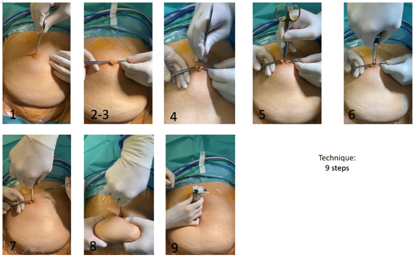
Entry-time assessment and historical comparator
Entry time in the robotic cohort was measured by the surgical team using a stopwatch. Timing began at the start of the skin incision and ended when the trocar had been completely inserted into the abdominal cavity. The recorded entry times therefore represented the time required for creation of the transumbilical access and complete trocar insertion. The mean entry time was 45 seconds, with an observed range of 40–65 seconds.
For contextual comparison, a historical cohort of 200 patients who underwent conventional laparoscopic surgery using Veress needle entry during the same study period was retrospectively reviewed.
The Veress cohort was not considered a directly comparable control group because the patients underwent different surgical procedures and had different gynecologic indications. The historical cohort was therefore used only to provide descriptive contextual information regarding the duration of abdominal entry.
Available demographic data showed broadly similar age and BMI distributions between the two cohorts. In the Veress cohort, 12% of patients were aged 35–44 years, 23% were aged 45–54 years, 35% were aged 55–64 years, 27% were aged 65–74 years, and 3% were aged 75–85 years.
Regarding BMI in the Veress cohort, 40.7% of patients had a BMI <25 kg/m², 42.3% had a BMI of 25–<30 kg/m², 11% had a BMI of 30–<35 kg/m², 4% had a BMI of 35–<40 kg/m², and 2% had a BMI ≥40 kg/m².
Previous abdominal surgery was documented in approximately 35% of patients in the Veress cohort. Previous hernia surgery was specifically documented in approximately 1%.
Because the Veress cohort was historical, represented different surgical indications, and was used solely for contextual comparison of entry time, no attempt was made to adjust for differences in surgical indication or other clinical characteristics.
Entry times in the Veress cohort were categorized as 3 to < 4 minutes, 4 to <5 minutes, and ≥5 minutes: 60 patients (30%) had an entry time of 3 to < 4 minutes, 120 patients (60%) had an entry time of 4 to <5 minutes, and 20 patients (10%) had an entry time of ≥5 minutes.
Because the Veress cohort represented a historical comparator and entry-time data were available as categorized rather than individual-level measurements, the comparison was descriptive. No inferential statistical test was performed to establish statistical superiority of either technique.
Postoperative follow-up
Postoperative follow-up was performed according to the standard clinical pathway associated with the underlying surgical indication.
All 110 patients undergoing robotic surgery for endometrial cancer were reassessed at our institution approximately 3 weeks after surgery. All 231 patients undergoing robotic-assisted surgery for pelvic organ prolapse were reassessed approximately 3 months postoperatively.
Among the 60 patients undergoing robotic surgery for benign gynecologic conditions, postoperative clinical assessment at our institution was also available for all patients included in the study 3-6 weeks after surgery. Thus, postoperative clinical documentation was available for all 401 patients included in the study cohort.
During the available postoperative assessments, the umbilical access site was evaluated for pain, infection, wound complications, hernia, or other access-related symptoms. No clinically relevant access-related postoperative problem was documented during the scheduled postoperative assessments.
Long-term systematic surveillance specifically for incisional or umbilical hernia was not performed.
The historical Veress cohort was not systematically assessed for postoperative umbilical outcomes because it was used solely as a descriptive comparator for entry time and postoperative follow-up was not part of the predefined comparative assessment.
Statistical analysis
Continuous variables are presented using descriptive statistics, and categorical variables are presented as absolute numbers and percentages.
Entry time in the robotic cohort is reported as mean and range. Entry-time data in the historical Veress cohort are reported as absolute numbers and percentages for each predefined time category.
No formal inferential statistical comparison was performed between the robotic and Veress cohorts. This decision was based on the historical nature of the comparator, differences in surgical indications and patient populations, and the different format in which entry-time data were available.
The analysis was therefore descriptive and was intended to characterize the feasibility and recorded entry time of the described technique rather than to demonstrate superiority over Veress needle entry.
A total of 401 patients undergoing robotic-assisted gynecologic surgery were included in the retrospective analysis.
The transumbilical eversion with direct trocar insertion technique was successfully completed in all 401 cases. No access failure was documented, and no conversion to an alternative abdominal entry technique was required.
The mean time from skin incision to complete trocar insertion was 45 seconds, with an observed range of 40–65 seconds. Entry time was measured and recorded by the surgical team using a stopwatch.
In the historical cohort of 200 patients undergoing conventional laparoscopy with Veress needle entry, entry time was 3 to < 4 minutes in 30% of patients, 4 to <5 minutes in 60%, and ≥5 minutes in 10%. These data are presented solely as a descriptive historical comparison.
No clinically relevant access-related complication was documented in the robotic cohort. Specifically, no vascular or visceral injury, clinically relevant entry-related bleeding, trocar malposition requiring corrective intervention, or other clinically relevant access-related intraoperative complication was observed.
No patient required laparotomy because of failure or complications of intraperitoneal access.
During the available postoperative follow-up, no umbilical wound infection, wound dehiscence, clinically relevant umbilical pain, or umbilical hernia related to the access site was documented.
All 110 patients with endometrial cancer underwent institutional postoperative assessment approximately 3 weeks after surgery. All 231 patients undergoing surgery for pelvic organ prolapse underwent assessment approximately 3 months postoperatively. All 60 patients undergoing surgery for benign gynecologic disease included in the analysis also had postoperative clinical documentation available at our institution with a follow-up visit 3-6 weeks after surgery.
No clinically relevant access-related postoperative problem was documented during these scheduled postoperative assessments.
Overall, the technique was successfully implemented throughout the study period without documented access failure or clinically relevant access-related complications in the analyzed cohort.
Safe and reliable abdominal entry remains a fundamental component of minimally invasive surgery. Despite the availability of several entry techniques, no single approach has been universally established as superior, and the choice of technique continues to depend on patient anatomy, surgical indication, and surgeon experience.1,2
The rationale for the technique described in the present study is based on the particular anatomy of the umbilical region. The umbilicus represents a natural anatomical landmark associated with the embryological development and closure of the anterior abdominal wall.6 Complete eversion allows the umbilical base to be directly exposed and elevated before creation of the entry opening. The trocar can subsequently be introduced along the vertical axis of the umbilicus.
The present technique is also designed to establish a perpendicular entry trajectory. The theoretical rationale is that a vertical trajectory may provide a short and controlled pathway through the umbilical region while the abdominal wall is elevated. However, these potential biomechanical advantages remain theoretical in the absence of direct biomechanical measurements, and the present clinical study does not establish that a 90° trajectory independently reduces tissue trauma or entry-related complications.
Previous reports have described techniques incorporating umbilical eversion and incision of the umbilical base. Sakamoto et al. described a modified closed-entry technique in which the umbilical ring was elevated with Kocher forceps, the umbilicus was completely everted, and the base was incised longitudinally.7 Their technique subsequently used a Step trocar to introduce a Veress needle, followed by pneumoperitoneum, removal of the inner needle, dilatation of the expandable sleeve, and final trocar placement. Thus, although the anatomical principle of complete umbilical eversion and base incision is similar, the access sequence differs substantially from the present technique, which creates the fascial and peritoneal opening directly with closed scissors and introduces the optical trocar immediately before pneumoperitoneum.
The anatomical concept underlying our approach also has some similarity to the technique described by Nikolopoulos et al., who reported an open entry technique using a congenital defect in the umbilical fascia.8 Their approach differs from the present technique because it represents a modification of open Hasson entry, whereas our method combines complete umbilical eversion with direct trocar insertion through a preformed transumbilical opening.
In the present cohort of 401 patients, the technique was successfully completed in all cases, with no documented access failures or clinically relevant access-related complications. The mean recorded entry time was 45 seconds, with an observed range of 40–65 seconds. These findings indicate that the technique was feasible within our institutional robotic surgery program and could be performed consistently by the six experienced gynecologic surgeons involved in the program.
The historical Veress comparator provides contextual information regarding entry time. Among 200 patients undergoing conventional laparoscopy during the same period, entry times were 3 to < 4 minutes in 30% of patients, 4 to <5 minutes in 60%, and ≥5 minutes in 10% of cases. These observations demonstrate a difference in the recorded duration of abdominal entry between the two cohorts. However, the Veress cohort was not a directly comparable control group because the patients underwent different surgical procedures and had different gynecologic indications. Furthermore, the historical nature of the comparator and differences in clinical workflow may have influenced the recorded entry times.
The Veress cohort was therefore included only to provide descriptive context for the duration of abdominal entry rather than to establish comparative efficacy or superiority. Although the available age and BMI distributions were broadly similar between the cohorts, demographic similarity does not eliminate potential differences in surgical complexity, indication, operative setting, or other unmeasured characteristics.
Previous evidence has not established a universally superior laparoscopic entry technique. The Cochrane review by Ahmad et al. concluded that the available evidence was insufficient to establish one entry technique as superior to another.1 Nishimura et al., in a systematic review and meta-analysis evaluating complications associated with three entry techniques, similarly found no statistically significant difference in major complications between direct trocar and Veress needle entry, while minor complications were less frequent with direct trocar entry.2 These findings support the consideration of direct trocar entry as an established alternative to Veress needle entry while emphasizing the limited frequency of serious entry-related complications.
The technique may be of particular practical interest in patients with obesity. Elevation and eversion of the umbilicus provide direct visualization of the umbilical base and permit the surgeon to establish the entry site before trocar insertion. In our cohort, 24.2% of patients had a BMI ≥30 kg/m², including 33 patients with BMI ≥35 kg/m² and 13 patients with BMI ≥40 kg/m². Nevertheless, the present study was not specifically designed or powered to evaluate outcomes according to BMI categories. Therefore, the findings should not be interpreted as demonstrating superior safety or efficacy specifically in patients with obesity.
Previous abdominal surgery was also not considered a contraindication in our institutional practice. A total of 170 patients (42.4%) had a history of previous abdominal surgery, including 10 patients with previous hernia surgery. The successful use of the technique in these patients indicates that previous abdominal surgery did not preclude its application in our cohort. However, previous umbilical surgery or scarring may alter local anatomy, and careful assessment of the umbilical region remains necessary before entry. The present study did not systematically evaluate the specific type or anatomical location of previous abdominal operations.
From a cosmetic perspective, the technique uses a median sagittal transumbilical incision. Previous work has evaluated different umbilical entry routes and has suggested that incision orientation may influence postoperative scar characteristics.9 However, cosmetic outcome was not a primary endpoint of the present study, and therefore no direct conclusion regarding a cosmetic advantage of the present technique can be drawn.
An important consideration is that direct trocar insertion involves passage into the abdominal cavity before establishment of pneumoperitoneum. As with all laparoscopic entry techniques, potential complications include vascular or visceral injury and bleeding. Umbilical eversion and abdominal wall elevation do not eliminate these risks. Appropriate patient assessment, controlled advancement of the instruments, knowledge of abdominal wall anatomy, and adequate surgical experience remain essential.
The absence of documented access-related complications in this cohort should also be interpreted in the context of the relatively low incidence of severe entry-related complications and the retrospective nature of the assessment. The present study was not designed to detect rare complications with sufficient statistical power, and the absence of documented events should not be interpreted as proof that the technique eliminates such risks.
The technique also has potential practical advantages. The standardized sequence consists of reproducible steps and does not require preliminary Veress needle placement. In our institutional experience, this allowed abdominal entry to be performed rapidly and according to a consistent protocol. However, the reproducibility observed in this study reflects the experience of an established robotic gynecologic surgery team and may not necessarily be transferable to surgeons without comparable experience.
Limitations
First, this was a retrospective, single-center observational study, which limits the generalizability of the findings.
Second, the study was conducted within an established robotic gynecologic surgery program and all entries were performed by experienced surgeons. Therefore, the results may not be directly applicable to less experienced surgeons or other surgical settings.
Third, the Veress comparator was historical rather than contemporaneous, and the two cohorts represented different surgical settings and surgical indications. Consequently, differences in workflow, operative complexity, patient selection, and measurement conditions may have influenced entry times. The Veress cohort was therefore used only as a descriptive contextual comparator and not as a matched control group.
Fourth, the study was not specifically designed to assess outcomes according to BMI, previous abdominal surgery, or individual surgical indication, and no dedicated subgroup analysis was performed. Long-term systematic surveillance specifically for incisional or umbilical hernia was not performed. Therefore, the absence of documented postoperative umbilical complications reflects the available clinical follow-up and should not be interpreted as evidence of absence of late complications.
Despite these limitations, the present study provides a detailed description of a standardized transumbilical entry technique and reports its implementation in a cohort of 401 patients within a specialized robotic gynecologic surgery program. Prospective multicenter studies with standardized outcome definitions, systematic follow-up, and a contemporaneous comparator group would be valuable to further evaluate the safety, efficiency, and generalizability of this approach.10
This study describes a standardized transumbilical entry technique combining complete umbilical eversion with direct trocar insertion for robotic-assisted gynecologic surgery. In this retrospective single-center cohort of 401 patients, the technique was successfully performed in all cases, with a mean entry time of 45 seconds and no documented clinically relevant access-related complications.
The findings suggest that this approach is feasible and reproducible within an experienced robotic gynecologic surgery team and may facilitate rapid establishment of abdominal access. Its use was feasible in patients with varying BMI and in patients with previous abdominal surgery, although the study was not specifically designed to establish differential safety or efficacy in these subgroups.
The historical Veress cohort was included only as a descriptive reference for entry time and was not considered a directly comparable control group because of differences in surgical indications and clinical setting. Accordingly, the present findings should not be interpreted as evidence of superiority of direct trocar insertion over Veress needle entry.
Given the retrospective design, single-center setting, experienced surgical team, and use of a historical rather than contemporaneous comparator, these findings should be interpreted as an initial clinical experience rather than definitive evidence of comparative safety or efficacy. Prospective comparative studies are warranted to further evaluate the safety, efficiency, and generalizability of umbilical eversion combined with direct trocar insertion
The continued support of the OR staff of Klinikum Nuremberg in providing state of the art patient care is greatly appreciated.
Ethics statement
This retrospective study was conducted using anonymized clinical data extracted from the institutional electronic medical records. According to the institutional assessment, approval by the ethics committee was not required for this retrospective analysis because the study used anonymized data and did not involve any intervention or modification of patient care.
The authors received funding from the Johann and Maria Künast Foundation to cover the article processing charges. The funder had no role in the study design, data collection, data analysis, interpretation of the results, manuscript preparation, or decision to submit the manuscript for publication.
Both Dimitrios Bolovis and Cosima Brucker hold a contract with Intuitive Surgical for proctoring and teaching using Da Vinci robotic systems. All authors declare that they have no other relevant financial or non-financial interests to disclose.

©2026 Lai, et al. This is an open access article distributed under the terms of the, which permits unrestricted use, distribution, and build upon your work non-commercially.