eISSN: 2377-4304


Research Article Volume 16 Issue 2
1Fellow literature, Obstetrics and Gynecology, Assiut University, Egypt
10Assistant Professor of Obstetrics and Gynecology, Aswan University, Egypt
11Lecturer of Obstetrics and Gynecology, Suez Canal University, Egypt
2Assistant Professor, Reproductive health and Family planning Department, National Research Centre, Egypt
3Assistant Professor, Obstetrics and Gynecology, Assiut University, Egypt
4Professor of Obstetrics and Gynecology, Assiut University, Egypt
5Professor of pediatric cardiology, Assiut University, Egypt
6Literature of obstetrics and Gynecology, Sohag University, Egypt
7Assistant professor of Obstetrics and Gynecology, Alazhar University, Egypt
8Lecturer of Obstetrics and Gynecology, Materia Teaching Hospital, Egypt
9Consultant of Obstetrics and Gynecology, Egypt
Correspondence: Mahmoud Alalfy, Reproductive health and family planning department, National Research Centre, Dokki, P.O:12622, Egypt, Tel +201002611058
Received: February 27, 2025 | Published: March 18, 2025
Citation: Ali S, Alalfy M, Ibrahim M, et al. Seif and Alalfy twist sign; a Novel view in fetal echocardiography in evaluation of normal heart. Obstet Gynecol Int J. 2025;16(2):47-50. DOI: 10.15406/ogij.2025.16.00787
Background: Fetal echocardiography is the primary tool for assessment of fetal heart and involves analysis of the cardiac anatomy from many views and Doppler examination of the intracardiac structures, great vessels, and umbilical artery.
Patients and methods: A prospective observational comparative study with comparing the Seif and Alalfy twist sign to the standard fetal echocardiography in evaluation of normal heart.
Results: Comparison of the Seif and Alalfy twist sign and fetal echocardiography in evaluation of the 4 chamber view revealed same findings.
Comparison between Seif and Alalfy twist sign and fetal echocardiography in assessment of outflow tracts revealed the same finding.
Conclusion: Seif and Alalfy twist sign is a reliable ultrasound (US) technique in scanning normal heart and could be compared to detailed fetal echocardiography.
Sief and Alalfy twist sign as a novel sign in fetal echocardiography as a new, easy, time effective view one step fetal heart examination.
Keywords: US, fetal, heart, echocardiography, Seif and Alalfy sign
US, ultrasound; LVOFT, left ventricular outflow tract; RVOFT, right ventricular outflow tract; 4CH V, 4 chamber view; 3VV, 3 vessel view; 3VTV, 3vessel tracheal view
Fetal echocardiography is the primary modality for assessment of fetal heart and needs detailed examination of the cardiac anatomy from many views and Doppler examination of the intracardiac structures and great vessels.1
Referrals for fetal echocardiography is dependent on maternal , fetal and familial risk factors ; but nearly fifty percent of neonates who have a congenital heart disease have no risk factor, and most of them were subjected to ultrasound during gestation that did not diagnose heart abnormality.2
Developments in US facilitates fetal heart examination in early pregnancy.3
Detailed cardiac scanning can be accomplished with the help of new US softwares.4,5 We aim to find a new, easy, time effective view one step fetal heart examination.
Our study describe Sief and Alalfy twist sign as a novel sign in fetal echocardiography as a new, easy, time effective view one step fetal heart examination.
Study design
A prospective observational study was performed pregnant women with age between 18- 40 years with no medical disorders pregnant between 18-26 weeks.
Study population
An observational study was conducted to investigates Sief and Alalfy Twist sign as novel sign in fetal heart examination in comparison with classic standard views.
Patients who gave a written consent to participate in the study was subjected to a uniform antenatal assessment protocol that includes basic obstetrics ultrasound examination, fetal anatomy scan & fetal echocardiography.
Criteria
Inclusion criteria: Singleton fetus, gestational age 18-26 weeks and no maternal medical disorder.
Exclusion criteria: Evidence of fetal infection, chorioamnionitis, fetal anomalies, abnormal fetal karyotype, patient withdrawal from the study and/or unavailability for follow-up.
Primary outcome measure
Obtaining clear images for fetal cardiac structures via standard views which are 4 chamber view (4CH V), outflow tracts and 3 vessel views.
Technique description
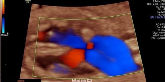
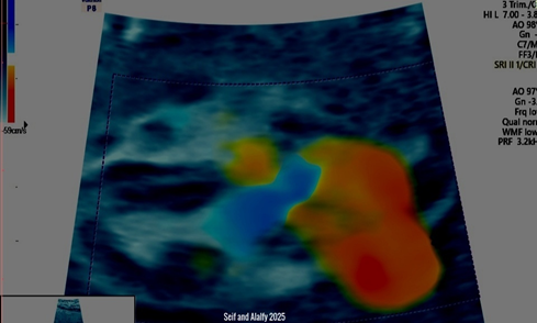
Seif and Alalfy twist sign, this technique describes an oblique view that illustrates the right and left ventricles with the interventricular septum and the atrioventricular valves with the aorta and pulmonary arteries crossing eachothers and till they form the v-sign ending to the ductus arteriosus as seen in Figure 1 and Figure 2.

Figure 1 Shows 2D US with Color Doppler illustrating Seif and Alalfy twist sign ,this technique describes an oblique view that illustrates the right and left ventricles with the interventricular septum and the atrioventricular valves with the aorta and pulmonary arteries crossing each others and till they form the v-sign ending to the ductus arteriosus.

Figure 2 Shows 3D US with Color Doppler illustrating Seif and Alalfy twist sign ,this technique describes an oblique view that illustrates the right and left ventricles with the interventricular septum and the atrioventricular valves with the aorta and pulmonary arteries crossing each others and till they form the v-sign ending to the ductus arteriosus.
This sign can be approached by sweeping the probe to get the optimum oblique view in the heart to illustrate right, left ventricles, interventricular septum, atrioventricular valves with the aorta and pulmonary arteries crossing each others.
The Seif and Alalfy twist sign can thus demonstrates a normal four chamber view and outflow tracts and 3 vessel views in a very short time Compared to the illustrated fetal echocardiography.
Comparison of the Seif and Alalfy twist sign and fetal echocardiography in evaluation of the 4 chamber view revealed same findings (Table 1) (Table 2).
|
POV |
Included patient (50) |
|
AGE |
26.44+ 4.24 |
|
BMI |
26.48+ 2.49 |
|
Gestational |
22.22+ 2.48 |
|
Parity |
|
|
Nullipara |
5 (10%) |
|
1 |
17 (34%) |
|
2 |
17 (34%) |
|
3 |
5 (10%) |
|
4 |
5 (10%) |
|
5 |
1 (2%) |
|
Gravity |
|
|
1 |
5 (10%) |
|
2 |
16 (32%) |
|
3 |
12 (24%) |
|
4 |
9 (18%) |
|
5 |
6 (12%) |
|
6 |
2 (4%) |
Table 1 Demonstrates baseline characteristics of women involved in the study
|
Screening |
Seif and Alalfy twist sign |
4 chambers by twist sign |
Outflow tract |
3 vessel view |
P-value |
|
Normal |
50 (100%) |
50 (100%) |
50 (100%) |
50 (100%) |
1 |
|
Abnormal |
0 |
0 |
0 |
0 |
Table 2 Shows the finding of the fetal heart scan by Seif and Alalfy twist sign and by detailed fetal heart scan; 4 chamber view, outflow tracts and 3vessel view
Comparison between Seif and Alalfy twist sign and outflow tract views in fetal echocardiography in assessment of outflow tracts revealed the same finding Seif and Alalfy twist sign, this technique could clearly visualize the 3 vessel view.
Statistical methods
Data were statistically described in terms of mean ± standard deviation (±SD), and range, or frequencies (number of cases) and percentages when appropriate. Correlation between various variables was done using Spearman rank correlation equation. .p values less than 0.05 was considered statistically significant.
All statistical calculations were done using the computerprogram IBM SPSS (Statistical Package for the SocialScience; IBM Corp, Armonk, NY, USA) release 22 for Microsoft Windows.
Situs and the four-chamber view
Sonographic technique: To determine cardiac situs, it is important to define fetal laterality, i.e. to recognize fetal left and right sides, with heart and stomach situated on the left side of the fetus.6–11
Four-chamber view: Evaluation of the four-chamber view involves careful scanning of special parts of right and left ariums and ventricles. A normal heart does not occupy more than one-third of the chest area.12–16
Seif and Alalfy twist sign, with its advantage as an oblique view helps to clearly identify the interventricular septum. Comparison of the Seif and Alalfy twist sign and fetal echocardiography in evaluation of the 4 chamber view revealed same findings.
Persistent bradycardia (≤110 bpm) in necessitates proper scan by a fetal cardiac specialist. Bradycardia could be due to frequent blocked atrial ectopic beats, atrioventricular block and sinusbradycardia. Persistent tachycardia (≥180 bpm has to be assessed for more serious tachydysrhythmias or fetal hypoxia.17–22
The ventricular septum must be scanned for defects from the apex to the crux.23,24
Comparison between Seif and Alalfy twist sign and outflow tract views in fetal echocardiography in assessment of outflow tracts revealed the same finding.
Outflow-tract, three-vessel and three-vessel-and-trachea views
The left ventricular outflow tract (LVOT) and right ventricular outflow tract (RVOT) views and the three-vessel (3VV) and three-vessel-and-trachea (3VTV) views are considered an essential part of the fetal cardiac examination.25
Seif and Alalfy twist sign is an oblique view that illustrates the right and left atriums and ventricles with the interventricular septum, so could help in properly evaluating the 4 chambers of the heart.
Seif and Alalfy twist sign, this technique could clearly visualize the 3 vessel view.
It is particularly useful for detection of aortic arch anomalies as aberrant right subclavian artery and coarctation of the aorta,vascular rings.26
Comparison between Seif and Alalfy twist sign and 3 vessels views in fetal echocardiography in assessment of 3 vessel view revealed the same finding.
The RVOT view settles the existence of a great vessel, the pulmonary artery, arising from the right and gives branches after a short passage. The pulmonary valve has a free movement and must not be have a thick wall.5
Left ventricular outflow-tract (LVOT) view
The LVOT view settles the existence of a great vessel arising from the left ventricle and from the center of the heart. Continuity must be illustrated between the ventricular septum and the anterior wall of aorta to prove integrity of the septum outlet arising from it.
Seif and Alalfy twist sign could evaluate the atrioventricular valves, outflow tracts with the aorta and pulmonary arteries crossing each others and till they form the v-sign ending to the ductus arteriosus.
To the best of our Knowledge, this is a novel sign that was not described previously inn the literature.
Seif and Alalfy twist sign is a reliable US technique in scanning normal heart and could be compared to detailed fetal echocardiography.
Sief and Alalfy twist sign as a novel sign in fetal echocardiography as a new, easy, time effective view one step fetal heart examination.
None.
None.
The authors declare that they have no competing interests.

©2025 Ali, et al. This is an open access article distributed under the terms of the, which permits unrestricted use, distribution, and build upon your work non-commercially.