MOJ
eISSN: 2379-6162


Case Report Volume 12 Issue 3
1Hospital Clínico Universitario de Valladolid, Spain
1Hospital Clínico Universitario de Valladolid, Spain
Correspondence: Marta Gallego Verdejo, Hospital Clínico Universitario de Valladolid, Spain
Received: September 30, 2024 | Published: December 17, 2024
Citation: Verdejo MG, Bailón FL, Pérez CR, et al. Radiological findings in orbital compartment syndrome. MOJ Surgery. 2024;12(3):129-130. DOI: 10.15406/mojs.2024.12.00279
Ocular compartment syndrome is a critical ophthalmological condition that demands immediate attention. If not promptly identified and treated, it can result in permanent vision loss. The condition arises when there is a sudden increase in the pressure within the orbit, often due to significant trauma that leads to substantial bleeding in this area. We will present the case of a 69-year-old patient who suffered an ocular compartment syndrome after trauma. We will also explain the main findings in CT that should make us suspect this entity, as well as the management that is usually carried out in these cases.1–3
We present the case of a 69-year-old male who came to the emergency department due to trauma to the right eye, after having been stabbed with a metal bar. There was no known ophthalmological or personal history of interest. Physical examination revealed a right periocular hematoma that did not allow the eyelid to open or move. In addition, a complete hyposphagma was observed, with a dull, intact cornea and a non-reactive right pupil. The eye fundus was not assessable during the examination.
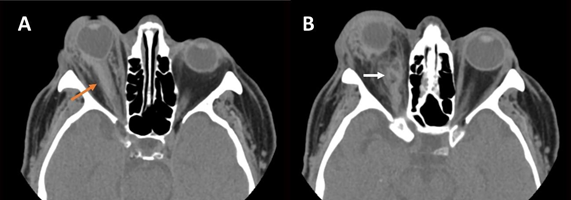
Given the findings, a CT scan of the orbits was decided upon. This revealed marked proptosis with deformity of the right eyeball, elongation of the optic nerve with perineural hematoma, as well as signs of retrobulbar hemorrhage. These findings were compatible with ocular compartment syndrome, so it was decided to perform an urgent canthotomy with septolysis. Unfortunately, the surgical treatment was not sufficient and the patient did not recover visual acuity, probably secondary to the damage suffered in the optic nerve.
Ocular compartment syndrome is considered an ophthalmological emergency, as it can lead to permanent vision loss if not identified in time. It occurs when the pressure inside the orbit increases suddenly. It is usually secondary to trauma that causes significant bleeding inside the orbit.
Clinically, patients present a decreased visual acuity, with a tense eyeball, increased intraocular pressure, proptosis, relative afferent pupillary defect, and restriction in eye movements. If intraocular pressure increases sufficiently, it can decrease perfusion of the retina and the optic nerve, leading to ischemia and permanent vision loss. The test of choice is CT, where proptosis of the affected eyeball is observed, as well as a deformity of its posterior pole with an angle less than 130º, known as the “guitar pick” sign. This angle results from two tangential lines, one to the medial wall of the eyeball and the other to the lateral wall, converging at the insertion of the optic nerve. If this angle is less than 120º it indicates severe proptosis with an unfavorable prognosis.4,5
Stretching of the optic nerve, a smaller caliber superior ophthalmic vein in the affected eye, and the presence of retrobulbar hemorrhage can also be observed. There are no significant differences in the size and shape of the extraocular muscles. Given its association with a history of trauma, patients may have intracranial hemorrhages and facial fractures. Likewise, ocular ultrasound can be useful in identifying signs that suggest the existence of an ocular compartment syndrome, such as the “guitar pick” sign, or the assessment of ocular vascularization by the Doppler study.6
The treatment is urgent lateral canthotomy or septolysis to reduce intraocular pressure. In cases where these are insufficient, orbital bone decompression can be performed. Despite treatment, if this pathology is not treated in time, it can lead to irreversible visual loss. For example, in the case series of Papadiochos et al.7 of the 9 patients diagnosed with ocular compartment syndrome, only 3 managed to preserve adequate visual acuity (Figure 1).

Figure 1 Ocular compartment syndrome secondary to trauma in the right eye. Axial sections of non-contrast-enhanced orbital CT showing A) marked right proptosis with deformity of the posterior pole known as “guitar pick” sign, as well as elongation of the ipsilateral optic nerve, with perineural hemorrhage (orange arrow). Image B shows retroocular hemorrhage in the medial aspect of the intraconal space (white arrow).
The role of the radiologist in ocular compartment syndrome is essential to identify the findings that indicate the presence of this pathology so that effective early surgical treatment can be achieved.
None.
The authors declare that there are no conflicts of interest.

©2024 Verdejo, et al. This is an open access article distributed under the terms of the, which permits unrestricted use, distribution, and build upon your work non-commercially.