MOJ
eISSN: 2379-6162


Case Report Volume 13 Issue 1
1Plastic and Reconstructive Surgery Unit, Hospital Regional de Alta Especialidad Ixtapaluca, Mexico
2Anesthesiology Unit, Hospital Regional de Alta Especialidad Ixtapaluca, Mexico
3Management Medical, Hospital Regional de Alta Especialidad Ixtapaluca, Mexico
4Investigation Unit, Hospital Regional de Alta Especialidad Ixtapaluca, Mexico
5Neurosurgery Unit Hospital Regional de Alta Especialidad Ixtapaluca, Mexico
Correspondence: Luis Licona Vite, Plastic and Reconstructive Surgery Unit, Hospital Regional de Alta Especialidad Ixtapaluca, Mexico
Received: April 01, 2025 | Published: April 17, 2025
Citation: Vite LL, Acosta GMS, Lopez GAG, et al. A new method for scalp avulsion treatment using dermal matrix and tnp negative pressure therapy: a clinical case report. MOJ Surg. 2025;13(1):30-33. DOI: 10.15406/mojs.2025.13.00289
This report corresponds to an intervention in a patient with a bone lesion of the scalp using sequential techniques, use of dermal matrix, followed by negative pressure therapy. The case stems from a 58-year-old woman who suffered an avulsion of the scalp after being caught by an agricultural machine, so she is referred to the Hospital Regional de Alta especialidad de Ixtapaluca, for surgical management. Joint surgery with neurosurgery was performed where initially a trephination was performed in “honeycomb” which functions as a vascularized medium to place the dermal matrix substitute and maintain angiogenesis, After this, it was covered with an autologous graft taken from the thigh 0.25mm thick. To increase tissue survival and decrease the risk of infections due to the extent of the lesion, used negative pressure therapy (TNP) with continuous pressure at 125 mmHg for 12 days. This treatment presented a notable regeneration of damaged tissues and decreased intrahospital stay time to 20 days as well as an adequate tissue integration, high survival of the tissues that revitalized the affected area.
Keywords: dermal matrix, scalp, reconstruction, avulsion, TNP
Avulsion of the scalp
The management of scalp avulsion lesions is a challenge for modern plastic surgery and not all patients are candidates for immediate tissue reimplantation.1 Scalp avulsion is not a common injury, it mainly occurs in work areas in which the worker does not wear protective equipment and in women with long hair who work in agricultural fields. These lesions can be so extensive that they can compromise the forehead area, eyebrows and up to the auricular area and can be so deep that it compromises the subgaleal area, damage to the periosteum and even present brain damage.2 Reconstruction in avulsion injuries in such extensive regions can be with autologous grafts, local flaps, and when it is feasible to reimplant the scalp in order to preserve the aesthetics.3
Tissue grafts
When they are not candidates for reimplantation, autologous grafts can be chosen but this requires a vascularized bed which is difficult, and slow to get, In such extensive trauma, one option is to opt for dermal matrix tissues developed by bioengineering. When a vascularized bed is needed, it is created from trephines in the diploe and the immediate placement of the dermal matrix tissues with which excellent results have been shown.4 Dermal matrix tissues can be used for this specific type of patients, futher provide cost accessibility and easy application. These are applied to recipient areas free of infection and are suitable in case of hypoxia. Types of bioengineered tissues are divided into natural tissues, artificial skin substitutes, cultured tissues and acellular dermal matrices.5
Within the Bioengineered tissues we highlight the substitutes of the dermis, that are easy to apply on a tissue that covers it (split thickness skin graft), its mechanism is not a re-epithelialization. Bioengineered dermal matrix tissues such as MatriDerm® provide a unique bovine collagen elastin matrix, serving as a dermal replacement scaffold. The dermal matrix is capable of integrating into the human dermis, accelerate cell invasion, the elongation, cell proliferation, limit the formation and contraction of myofibroblasts.6 In reconstruction therapy with bioengineered tissues, revascularization is a critical point and several factors can alter it, the most common being the formation of a seroma and/or hematoma that compromises tissue survival.7–10
Negative pressure therapy
At present, negative pressure methods are widely used in all types of wounds and their application in reconstruction with skin grafts it has been reported that it accelerates the time of tissue incorporation and reduces the negative factors for their integration.11–13 Vacuum systems (P. Example type TNP) has the following benefits: promotion of angiogenesis, the stimulation and formation of healing granulation tissue, as well as an increase in local blood flow in the wound area, helps to keep the graft firmly in place, reducing the formation of seromas or hematomas under the bandage and removes exudate to prevent maceration of skin grafts under the bandage, thus maintaining an environment that favors tissue integration and in our experience reduces tissue integration time to less than 50%.14,15
The following clinical case is presented, which corresponds to a 58-year-old female patient from the state of Puebla, resident of Ixtapaluca Estado de México, who suffered an avulsion of the scalp while working with an agricultural machine. (husker). She suffered an accident in the workplace, with craniocephalic trauma generating detachment of the hairy skin, and is transferred to the Hospital Regional Emilio Sánchez Piedras Tlaxcala, where cranial trephines were performed as a reconstructive surgical treatment method to generate granulation tissue in the biparietal area with bleeding and moderate pulsatile activity, as well as loss of 50% of the pinna, skin traction was performed to avoid retraction and loss of 80% of the scalp skin, transfer is made to the Hospital Regional de Alta Especialidad de Ixtapaluca (HRAEI) for assessment and continuation of medical-surgical care (Figure 1).
Clinical history and surgical procedure
Upon admission to the HRAEI the female patient. Not responding to external stimuli, aphasica normocephaly with total scalp loss (Figure 1) by avulsion covered with sterile bandage, normoreflexic isochoric pupils, of 3 mm approximately, photomotor and consensual reflex, muscle stretching reflexes, without focus data, cardiopulmonary without commitment, abdomen without alterations, symmetrical extremities, eutrophics, pulses present in all extremities and capillary filling 2 seconds.
Therapeutic intervention
The patient is admitted to the operating room in a joint surgery by the plastic surgery and neurosurgery services. In the first half of the first stage, it began with antisepsis and asepsis of the surgical site, followed by scalp trichotomy with surgical cleansing with wound solution (Vashe).
Multicranial trephine neurosurgical repair
Start trepanation or generation of multiple trephines with a midas type craniotome rex using a pediatric sized self-locking starter drill with a deep limit of only reaching the diploe, which is identified by the venous bleeding obtained, this procedure is performed on both parietal bones without scalp skin, are made in the form of honeycombs (Figure 2) having a 5 mm spacing between each one.
Once the multiple trephines have been completed, the cortical bone is scraped with a 3 mm diamond bur to remove necrotic tissue. After this procedure, the area is prepared for the placement of the dermal matrix tissues developed by bioengineering for which we use irrigation with wound solution and placement of gauze impregnated with the same solution for 5 minutes.
Placement of dermal matrix tissue
Gauze impregnated with antiseptic solution is removed. Dermal matrix is placed (MatriDerm®) of 1mm on the exposed bone and the multiple trephines made without any fixation system, irrigated with saline solution to promote immediate adhesion (Figure 3).
Application of skin graft - A 0.25 mm partial thickness skin graft obtained from the right pelvic limb is taken for direct application on the dermal matrix and fixed with staples (Figure 4).
System placement TNP
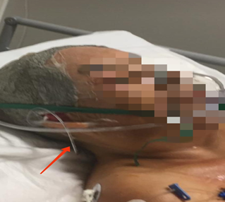
Dressing was applied UrgoTul of Lipid-Colloidal technology and sponge application for vacuum-assisted wound treatment by vacuum closure T.N.P. covering outside of the perilesional area (4 cm) and is fixed with Steri-Drape then install a negative pressure machine type TNP, we proceed to verify absence of leaks we program the system to 125 mmHg of negative pressure, with continuous pressure, low. This procedure was carried out during 6 days (Figure 5).

Figure 5 Functional sealing of the system TNP which include pinna (red arrow points to power supply probe).
Management with negative pressure therapy
After the surgical interventions by combining dermal matrix, grafting and TNP, to the 6 days we presented tissue preservation without loss of the anchoring matrix or grafts, and without signs of infection. The specific recommendation is that the system should be continuous throughout the 6 days, as well as the constant review of the system's programming. It was necessary for proper sealing of the system TNP to include the auricles, as well as protecting the ear canals with a 1 cm cotton swab inside the ear canal and a 3 mm feeding tube, which allowed us to maintain the function and protection of the ear canals (The patient, always had hearing pre and post negative pressure treatment).
The system replacement TNP was performed on the sixth day, without presenting any complications and obtaining total incorporation in the tissues, the system was removed TNP definitively at 12 days postoperatively, after removal, the tissues were left exposed. No other surgical or revision treatment was required (Figure 6).
Monitoring and results
After your discharge, was cited an external consultation for review in a month, where we observed the complete re-epithelialization of the graft area, no areas of necrosis, with an adequate integration of the tissues of the dermal matrix, with a 2 second capillary refill (Figure 7).
Although bioengineered dermal tissues began to be investigated in the 1960s, in our country they were used for the first time in clinical experimentation in the 1980s. At present, it has been insufficiently used, when its potential is very high in terms of success and profitability, the description of this article and case report is intended to establish a clear and concrete protocol for successful management of complex pericranial injuries with the support of bioengineered dermal matrix tissues, supported by other wound dressings such as TPN, the sum of this has been successful, and in the future new tissues will be developed that will facilitate the reconstruction of areas with larger lesions.
The problem of extensive lesions of pericranial tissues are and have been a challenge for surgeons, throughout history with high morbimortality and hospital stays, using dermal matrix tissues developed by bioengineering along with the available methods, has allowed us to manage and improve a case that was expected to reach term in months reducing hospital stay to days without associated morbimortality.
The technique and use of dermal matrix in combination with the therapy TNP, proved to be an efficient methodology in scalp avulsion addressed at the HRAEI., with a decrease in final hospital stay time to 20 days when alternative treatments average 2 to 4 months. In addition, a marked regeneration was demonstrated for this injury, with 80% regeneration of the scalp trauma compared to other techniques, which is efficient and allows the patient to heal without infection, without the formation of seromas or necrotic tissue, which commonly affects this type of injury.
None.
The authors declare that there are no conflicts of interest.