MOJ
eISSN: 2374-6939


Case Report Volume 17 Issue 3
1School of Medicine, National Defense Medical Center, Taipei, Taiwan
2Department of General Medicine, Tri-Service General Hospital, National Defense Medical Center, Taiwan
3Departments of Administration and Physical Therapy and Rehabilitation, Catholic Fuan Hospital, Dounan Township, Taiwan
4Department of Physical Medicine and Rehabilitation, Kaohsiung Veterans General Hospital, Taiwan
5Department of Physical Medicine and Rehabilitation, Tri-Service General Hospital, School of Medicine, National Defense Medical Center, Taiwan
Correspondence: Shin-Tsu Chang, Department of Physical Medicine and Rehabilitation, Kaohsiung Veterans General Hospital, Taiwan
Received: May 05, 2025 | Published: May 22, 2025
Citation: Kuan-Chieh W, Shin-Tsu C. Remission of pain post steroid injection at the pseudo-joint of transitional vertebra in a case of Bertolotti syndrome. MOJ Orthop Rheumatol. 2025;17(2):46-48. DOI: 10.15406/mojor.2025.17.00698
Bertolotti Syndrome, one of the reasons for lower back pain, is defined by transitional vertebra, scoliosis, and sciatica, and often treated with conservative therapy or rare surgical resection. We present a case of Bertolotti Syndrome with intermittent and recurring low back pain. The pain, observed as a distinct pseudo-joint, was identified by skeletal scintigraphy, showing significant therapeutic success without recurrence after a steroid injection. In the presence of Bertolotti Syndrome, skeletal scintigraphy, pinpointing the high intensity of pain localization, would be a feasible diagnostic approach.
Keywords: lumbosacral transitional vertebra (LSTV), Bertolotti syndrome, single-photon emission computed tomography, lower back pain, steroid, scintigraphic rehabilitation
Bertolotti Syndrome, one of the reasons for chronic lower back pain, is characterized by lumbosacral transitional vertebra (LSTV), scoliosis, and sciatica. LSTV is a type of congenital abnormality involving the fifth lumbar vertebra (L5) or the first sacral segment (S1).1 Recently, Jenkins proposed a new classification, based on a shift from transverse process (TP) size to the TP-ala gap as a metric reflection to supplement the deficiencies of Castellvi, which has been utilized with LSTV for a long time.2,3 Nevertheless, the unclassified transitional vertebra associated with Bertolotti Syndrome has piqued our interest, especially the presence of a large transverse process with a pseudo-joint at the other side. Unexpectedly, whether this anatomical site is a genuine source of pain is still under debate. Additionally, the efficacy of medication administration at this site for symptom relief is also a particular emphasis for us.
Single-photon emission computed tomography (SPECT) is a nuclear medicine imaging modality employed for the functional assessment of tissues and organs, distinguishing itself from purely anatomical imaging techniques.4 Furthermore, integrating images of SPECT and computed tomography (CT) provides explicit sites due to CT’s morphological characterization and more accurate localization from SPECT at the uptake site.5 To our amazement, SPECT can also play a crucial role not only in radiation treatment planning but also in the evaluation of therapy response.6 SPECT-CT was also employed to precisely identify the specific location and target the lesion for accurate therapeutic intervention in our case. We present a case with remarkably intense uptake in the pseudo-joint observed through scintigraphic imaging, demonstrating complete remission of pain following interventional steroid injections.
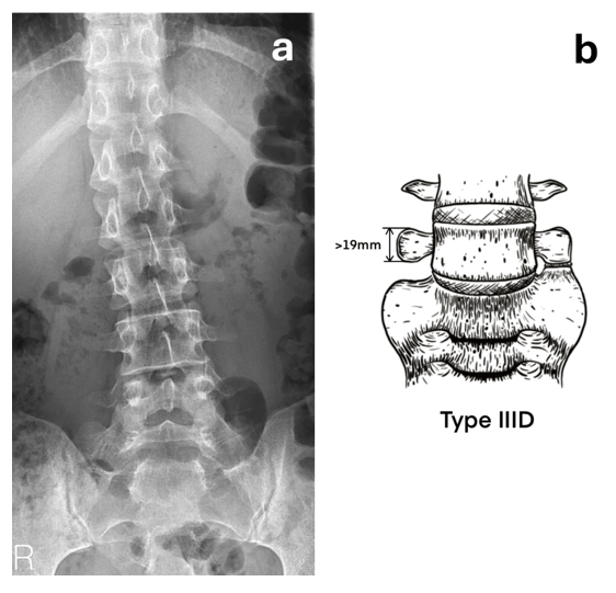
A 25-year-old male visited the rehabilitation outpatient department due to intermittent lower back tightness and sciatica for 2-3 years, correlated with participation in strenuous physical activities and recurring as before. Despite seeking help at a rehabilitation clinic and undergoing rehabilitation therapy, the effectiveness in alleviating symptoms was limited. Upon physical examination, the Fortin finger sign was identified on the left side, and X-ray assessment revealed minor lumbar spine scoliosis along with a transitional vertebra at the lumbosacral junction (Figure 1). Therefore, Bertolotti Syndrome was confirmed. Subsequently, Technetium-99m labelled methylene diphosphonate (Tc99m-MDP) SPECT-CT imaging was arranged to confirm the location of the lesion, especially the pseudo-joint.

Figure 1 (a) X-ray film of lumbar spine showing scoliosis and a transitional vertebra in the lumbosacral junction. (b) Compatible with our classification Type IIID, which show transverse process that measures 19 mm or more in height with a pseudo-joint on the opposite side.
Notably, SPECT revealed an exceptionally intense uptake in the left pseudo-joint, indicating the potential location of the severe pain (Figure 2). Eventually, after obtaining his agreement, significant efficacy was observed following steroid injection of the pseudo-joint. He did not visit our clinic in the subsequent two year.
We present a case of fitful and recurring lower back tenderness. Concurrently, X-ray revealed a typical transitional vertebra in the lumbosacral junction. Furthermore, SPECT-CT effectively showed intense uptake in the left pseudo-joint of this LSTV, indicating the location of the severe pain of the patient. Steroid injection was administered promptly and demonstrated significant therapeutic efficacy.
The classification of LSTV is crucial for a clear understanding and appropriate management of lower back pain condition. In addition to Tini classification,7 Castellvi Classification is also recognized as the most widely used system. In the reported case, we opted for a novel classification system over the traditional one, as the Castellvi classification fails to categorize certain instances of LSTV. The gap was addressed by Jenkins et al,2 focusing on the anatomical classification of LSTV variants by adding two more subtypes.
SPECT-CT distinguishes itself from purely anatomical imaging modalities by producing a 3-dimensional image of the distribution of a radioactive tracer.4 Additionally, degenerative diseases and neoplastic processes, especially inflammation in the musculoskeletal region, were all precisely detected by SPECT-CT.8,9 One case report even revealed that the pain improvement of surgical fusion was due to the successful evaluation of increased osteoblastic activity with anatomic precision by SPECT-CT.10 In our case, we implemented the identical methodology, achieving precise localization at the pseudo-joint. By administering steroid injections directly at the lesion, we achieved remarkable therapeutic outcomes. Given its efficacy in pain management, this procedure merits widespread adoption.
Some selected cases initially failed with steroid injection11 suggesting that treatment effectiveness may not necessarily be consistent across all types of Bertolotti Syndrome. Our patient, however, showed a distinct but excellent response. Interestingly, although one case used SPECT as a diagnostic tool11 similar to ours, its pattern was classified as type IIA, which does not align with any of the classifications defined by Castellvi.
Jenkins et al.2 proposed a novel classification in 2023 based on a shift from TP size to the TP-ala gap as a metric reflection, serving as a pivotal tool in the precise diagnosis and management of Bertolotti Syndrome. This refined classification aids in tailoring treatment approaches,12 ensuring more targeted and effective management of patients with these specific LSTV features. In contrast, steroid injection was employed in our case rather than Jenkin's recommendation of surgical resection or decompression of the pseudo joint (false joint). Nonetheless, our approach of steroid injection yielded a favorable outcome.
Our opinion diverges from Jenkins, particularly due to its shortcomings regarding the lack of a standardized and consistent approach to measuring the 10mm gap. This inconsistency also blurs the distinction between defined Types IIA and IIC, as they do not exhibit significant enlargement of the transverse process, making them appear almost identical when viewed through the Castellvi classification. Consequently, we propose a novel classification method as detailed below. Historically, we have shown a preference for the Tini classification,7 which highlights the symmetry of the transverse process. Type III in the Tini classification addresses asymmetric transverse processes, with Type III A featuring one side with a pseudo-joint and the other fused, Type III B featuring one side with a diminished transverse process and the other fused, and Type III C featuring one side with a diminished transverse process and the other with a pseudo-joint. However, it lacks representation of unilateral prominence of a large transverse process with a pseudo-joint on the opposite side.
In addition, we have introduced Type IVB, a category that has not been previously described in the literature. Chang et al,13 as early as 2008, identified various classifications of spina bifida occulta (SBO), including cases where the entire sacrum is disrupted. Indeed, the lumbar multifidus muscle, a critical component of the back extensor group and a deep stabilizer of the trunk, has an attachment site that coincides precisely with the location of the SBO defect. The presence of SBO may compromise the structural integrity of the lumbar multifidus muscle, potentially leading to postural instability, particularly under conditions of repeated mechanical stress. This instability may further progress to spondylolysis. Moreover, a direct correlation between the extent of the bony defect and the severity of proprioceptive deficits has been observed, highlighting the clinical importance of this classification.
In cases of Bertolotti Syndrome, Tc99m-MDP SPECT-CT, pinpointing the high intensity of pain localization, would be a feasible diagnostic approach.
None.
The authors declare that there are no conflicts of interest.

©2025 Kuan-Chieh, et al. This is an open access article distributed under the terms of the, which permits unrestricted use, distribution, and build upon your work non-commercially.