MOJ
eISSN: 2374-6939


Research Article Volume 18 Issue 3
 Vadim V Verbov, Ievgen V Cheshuk, Vitalii I Fortunenko
Department of spinal neurosurgery, Romodanov Neurosurgery Institute of National Academy of Medical Sciences of Ukraine, Ukraine
Correspondence: Oleksii S. Nekhlopochyn, Department of spinal neurosurgery, Romodanov Neurosurgery Institute of National Academy of Medical Sciences of Ukraine, Ukraine
Received: May 04, 2026 | Published: June 25, 2026
Citation: Nekhlopochyns OS, Verbov VV, Cheshuk IV, et al. Development and retrospective decision-level evaluation of an individualized treatment-planning algorithm for traumatic thoracolumbar junction injuries. MOJ Orthop Rheumatol. 2026;18(3):109-118. DOI: 10.15406/mojor.2026.18.00749
Background: Traumatic injuries of the thoracolumbar junction (Th11–L2) are one of the most common sites of spinal trauma and frequently combine structural instability with neurological deficit, while treatment planning - conservative versus operative management, short versus long fixation, decompression, anterior column reconstruction - varies substantially between surgeons. Classification systems describe injury severity but provide little formalization of the tactical extent of intervention. The aim was to present an individualized treatment-planning algorithm and to evaluate how observed structural failures mapped to predefined decision levels in a retrospective cohort.
Methods. The algorithm was assembled by integrating morphometric, prognostic, and biomechanical models and structured as if–then sequences separating the strategic, tactical, and technical decision levels. Its inputs are the AO Spine type, the morpho-functional variant, the neurological status by ISNCSCI (AIS grade), the spinal canal state, the posterior ligamentous complex (PLC) state, bone density (HU), mechanical exposure, and clinical context. The retrospective evaluation was performed on 326 patients (12-month follow-up in 297), matching endpoint events to the algorithm's branch points.
Results. The algorithm operates as a sequence of gates: a verification gate, a morphological branch (A1–A2, A3–A4, B, C), and neurological, ligamentous, densitometric, and contextual modifying gates, followed by the choice of strategy and tactical extent. Among the 297 patients with 12-month follow-up, 43 endpoint events occurred (41 structural failures and 2 clinico-deformational failures; an adverse outcome in 38 patients); 40 of the 41 structural failures mapped to the tactical level, and no isolated strategic-level failures were found. Corpectomy was associated with the numerically highest observed rate of structural failure (6/23; 26.1%; 95% CI 10.2–48.4%), all due to cage subsidence; this estimate was descriptive owing to the small subgroup size and probable confounding by indication, with no descriptive advantage in neurological recovery (47.8% versus 51.2%). The evaluation showed that the algorithm mainly refines the tactical rather than the strategic level of a decision.
Conclusions. The proposed algorithm provides a transparent framework for treatment planning, and its underlying modules showed internal performance compatible with exploratory risk stratification. In retrospective decision-level evaluation, most structural failures mapped to tactical branch points. The algorithm should be considered a decision-support tool and requires prospective external validation.
Keywords: thoracolumbar junction, spinal injury, treatment planning, clinical decision support, AO Spine, transpedicular fixation
Traumatic injuries of the thoracolumbar junction (Th11–L2) are one of the most common sites of spinal trauma and are frequently accompanied by structural instability and neurological deficit. The choice of treatment tactics remains debated: decisions on conservative versus operative management, short versus long transpedicular fixation (TPF), the need for decompression, and anterior column reconstruction vary substantially between surgeons and institutions.1
Current classification systems - the AO Spine classification2–4 and the TLICS severity score5 - standardize the description of injury morphology and guide the indication for intervention, yet they largely leave the choice of the tactical extent to the surgeon. The quantitative morphometric,6 densitometric (HU), biomechanical, and prognostic data accumulated for this region have not been integrated into a single operational framework for tactical decision-making, which sustains variability and complicates the retrospective analysis of sources of structural failure.
An adverse treatment outcome may originate from different decision levels: an erroneous strategic direction (e.g., withholding needed stabilization), an insufficient tactical extent (short fixation where long is required; a missed decompression), or a technical execution error. Classification scales do not separate these levels, which hampers both the retrospective analysis of structural failure and rational planning. Progression of post-traumatic kyphotic deformity, secondary stenosis, and loss of correction remain frequent causes of reoperation,7,8 underscoring the need for a transparent, reproducible sequence of tactical choice.
The aim of this work was to present an individualized treatment-planning algorithm for thoracolumbar junction injuries that sequentially translates morphological, neurological, morphometric, densitometric, biomechanical, and clinical-contextual features into a specific tactical zone, and to evaluate how observed structural failures mapped to predefined decision levels in a retrospective cohort.
Study design and cohort
The study was approved by the ethics and bioethics committee of the State Institution “Romodanov Neurosurgery Institute of the National Academy of Medical Sciences of Ukraine” (protocol No. 4 of 05 September 2018) and was performed as part of a planned research project (state registration number 0119U000110). For the prospective part, informed consent for the collection, processing, and publication of aggregated results with confidentiality was obtained from patients; the retrospective part was performed using de-identified clinical data in accordance with the ethics committee decision.
The study is based on a single-center retrospective-prospective cohort of patients aged ≥ 18 years with acute and subacute traumatic injuries of the thoracolumbar junction (Th11–L2). Exclusion criteria were severe combined trauma precluding adequate initial assessment; multiple spinal injuries with a dominant level outside Th11–L2; old consolidated injuries (> 6 months); marked degenerative changes or sequelae of prior surgery precluding correct morphometry; pathological, tumor, and infectious processes; and isolated osteoporotic insufficiency fractures without signs of structural traumatic disruption. The leading inclusion criterion was the injury morphology on imaging rather than reduced bone density alone. Analytical subsamples were formed from the main cohort according to each stage's data-completeness requirements: morphometric analysis by AO Spine type (A1–A2 - 57, A3–A4 - 100, B2 - 118, C - 45 cases), prediction of PLC injury with a verified reference state (90 cases), and the treatment-outcome analysis with a 12-month follow-up (297 patients). Patients were treated between January 2011 and May 2025. For morpho-functional modeling, type B analysis focused on the B2 subgroup (118 cases), whereas the treatment-outcome analysis included all type B injuries (124 cases). The baseline characteristics of the cohort are summarized in Table 1.
|
Characteristic |
Value (n = 326) |
|
Age, years, median [IQR] |
53 [31–65] (range 18–92) |
|
Sex — male / female, n (%) |
165 (50.6) / 161 (49.4) |
|
Treatment period |
January 2011 – May 2025 |
|
12-month follow-up, n (%) |
297 (91.1) |
|
AO Spine type — A1–A2 / A3–A4 / B / C, n |
57 / 100 / 124 / 45 |
|
AO Spine type — A1–A2 / A3–A4 / B / C, % |
17.5 / 30.7 / 38.0 / 13.8 |
|
Injury level — Th11 / Th12 / L1 / L2, % |
3.7 / 22.1 / 35.0 / 16.9 |
|
Injury level — multilevel / contiguous-level, % |
22.4 |
|
Baseline AIS — A / B / C / D / E, n |
72 / 56 / 59 / 75 / 64 |
|
Baseline AIS — A / B / C / D / E, % |
22.1 / 17.2 / 18.1 / 23.0 / 19.6 |
|
Native HU, median [IQR]; range |
183.6 [149–229]; 82–330; available in 311 (95.4%) |
|
Posterior edge distraction (PED), mm, median [IQR] |
3.9 [1.8–6.5] (n = 218) |
|
Treatment — conservative / cement augmentation, n |
13 / 41 |
|
Treatment — short TPF / long TPF without decompression, n |
54 / 19 |
|
Treatment — long TPF + decompression / corpectomy, n |
176 / 23 |
Table 1 Baseline characteristics of the cohort
Clinical, neurological, and radiological assessment
Injury morphology was classified using the AO Spine system. Neurological status was assessed on admission according to the International Standards for Neurological Classification of Spinal Cord Injury (ISNCSCI), with determination of the impairment grade on the AIS scale (ASIA Impairment Scale; A–E);9,10 where primary documentation used the Frankel scale, standardized recoding to AIS was performed for category-level analysis only, based on documented motor and sensory findings where available, with uncertain cases reviewed by two investigators. Radiological examination comprised two-projection plain radiography, spiral computed tomography (CT) with multiplanar reconstructions, and magnetic resonance imaging (MRI). CT was the main quantitative method, providing morphometry and measurement of trabecular bone density in Hounsfield units (HU).11,12 Native HU was measured in the cancellous bone of a vertebral body outside the injury zone; when the injured vertebral body was unsuitable for measurement, an adjacent intact vertebra was used. Native HU was available in 311 of the 326 patients (median 183.6; range 82–330). The region of interest was placed within trabecular bone, avoiding cortical bone, fracture lines, venous channels, focal sclerosis, and cement or metal artifacts. All morphometric measurements (Cobb and Gardner angles, A/P and AVH indices, anterior and posterior edge distraction AED and PED, rotation) were performed in RadiAnt DICOM Viewer by three independent experts blinded to clinical data and to the verified PLC state; the mean of three measurements was used for quantitative parameters. The PLC state was verified from a combination of MRI and CT signs, with MRI as the reference standard for the soft-tissue component.
Algorithm development
The algorithm is an integrative synthesis of five sequential analytical stages performed on the described cohort. (1) The morpho-functional typology within each AO Spine type was obtained by cluster analysis (k-means and Gaussian mixtures) of standardized CT morphometric parameters with preceding principal component analysis; on this basis, injury variants were defined - compensated / subcompensated / decompensated for A1–A2, kyphotic-deformational / compression-retropulsive for A3–A4, deformational / channel-compressing / mixed for B2, and single-plane / multi-plane / gross-destructive for C - along with operational threshold values approximated from the cluster boundaries. (2) The risk of PLC injury was predicted using separate gradient-boosting (XGBoost) models for A1–A2 and A3–A4, converted into point scales, nomograms, and risk categories.13–16 (3) The biomechanical justification of short and long transpedicular constructs, M1–M4 configurations, and corpectomy variants was performed using the finite element method on a three-dimensional Th9–L5 model under five loading modes (compression, flexion, extension, lateroflexion, rotation). (4) The efficacy of indirect decompression was assessed using a prognostic model of residual canal stenosis based on the initial stenosis and the F. Magerl morphological type.17 (5) The prognosis of clinical outcomes - including a nomogram of kyphotic-deformity progression risk - was obtained using regression models;18 neurological recovery was stratified separately according to baseline AIS grade and injury morphology.
The results of these stages were integrated into a hierarchical if–then algorithm separating three decision levels: the strategic level (whether intervention is needed and its main goal - observation, cement augmentation, stabilization, decompression, or anterior column reconstruction), the tactical level (the extent of intervention: short versus long TPF, the number and type of screws, intermediate screws, cross-links, augmentation, corpectomy), and the technical level (the quality of the surgical execution itself, deliberately excluded from modeling). The decision sequence is implemented through gates - verification, morphological, neurological, ligamentous, densitometric, and contextual. The input variables are divided into mandatory ones (AO Spine type, morpho-functional variant, AIS grade, spinal canal state, PLC state, HU, general clinical context) and modifying ones (mechanical exposure, age, pain, expected mobility, polytrauma, comorbidity); each variable is interpreted within a specific branch.
The models underlying the algorithm had the following internal-validation characteristics. The PLC injury prediction models were built using gradient boosting (XGBoost) separately for A1–A2 (n = 43) and A3–A4 (n = 47); from 17 initial CT parameters, 12 predictors were selected on the basis of a priori criteria of anatomical independence and measurement reproducibility (rather than on the target variable), so the reported AUC values are cross-validation estimates rather than in-sample ones. Under repeated stratified cross-validation, the AUC was 0.836 for A1–A2 and 0.894 for A3–A4, and the derived point models (logistic regression of the total score) reached AUC 0.944 and 0.880 at acceptable calibration (Brier score 0.103 for A3–A4). The kyphotic-deformity progression model was built with LASSO predictor selection and internal bootstrap validation per F. Harrell (B = 2000 resamples): apparent AUC = 0.907 at a mean optimism of 0.052. The prediction error of residual stenosis under indirect decompression was estimated as MAE 3.2 and RMSE 3.8 percentage points. The interobserver reproducibility of morphometry was high: the mean absolute error for angular parameters (Cobb, Gardner, rotation) was 0.4–0.7° (relative 2.9–4.0%), and for the A/P and AVH indices about 0.015 (2.2–2.7%). Owing to the limited size of the training subsamples, the PLC prognostic models should be regarded as tools of internally validated risk stratification rather than finally validated predictors. They were not used as externally validated predictors of outcome but as internally validated risk-stratification modules that define working decision-support thresholds; accordingly, all quantitative thresholds have the status of working reference points and should not be interpreted as automatic indications for surgery.
Retrospective evaluation
The retrospective evaluation of the algorithm was performed on the treatment-outcome subsample - 326 inpatients, of whom 297 had a 12-month follow-up (within 11–14 months). Endpoint events were predefined structural failure and clinico-deformational failure; loss of correction was defined as a recurrence of kyphotic deformity exceeding 5° from the postoperative value; the endpoint definitions are summarized in Table 2. The decision-level failure category (strategic, tactical, or technical) was assigned by two independent experts against predefined criteria (Table 3), and disagreements were resolved by consensus with a third expert. Cohen’s κ was not calculated because almost all events fell into a single (tactical) category, which makes the coefficient unstable and uninformative. Each event was matched to a decision-level failure category and to the corresponding algorithm branch point: a discrepancy between the baseline treatment direction and the injury morphology was assigned to the strategic level, an insufficient scope or configuration of intervention under a correct strategy to the tactical level, and an execution error to the technical level; assignment was based on the structural-failure pattern and the documented surgical decision. The evaluation aimed to verify logical correspondence rather than to prove prospective efficacy; interpretation was asymmetric - an adverse outcome was regarded as a marker of tactical insufficiency, whereas a favorable outcome was not taken as proof of optimal tactics.
|
Endpoint |
Definition |
Unit of analysis |
|
Loss of correction |
Recurrence of kyphotic deformity > 5° (Cobb angle) from the immediate postoperative value; > 10° regarded as clinically significant |
event |
|
Cage subsidence |
Subsidence or migration of the interbody cage ≥ 3 mm |
event |
|
Implant failure |
Screw loosening (peri-implant radiolucency > 2 mm), pull-out, screw or rod breakage, or malposition |
event |
|
Secondary stenosis |
Residual or secondary spinal canal compromise of clinical or radiological relevance |
event |
|
Neurological deterioration |
Decrease of ≥ 1 AIS grade or a new motor, sensory, or radicular deficit |
event |
|
Predefined structural failure |
Any of the above radiological, implant-associated, or canal-related events (excluding isolated neurological deterioration) |
event |
|
Clinico-deformational failure |
Pain and disability with deformity progression not meeting the structural-failure criteria |
patient/event |
|
Generalized adverse outcome |
Patient-level composite of an unsatisfactory clinico-radiological result (loss of correction > 10°, pseudarthrosis, implant migration/breakage, residual or secondary stenosis, NRS ≥ 7, AIS worsening, or reoperation) |
patient |
Table 2 Definitions of endpoint events
Note: Neurological deterioration was included in the generalized adverse outcome but was not counted as a structural failure unless accompanied by a structural substrate.
|
Decision level |
Definition |
Example |
Distinguished from |
|
Strategic-level failure |
Inappropriate baseline treatment direction despite available morphology and neurological data |
Conservative management despite a clearly unstable B/C injury |
technical-level failure |
|
Tactical-level failure |
Correct general strategy but insufficient extent or configuration of treatment |
Short TPF where long TPF is indicated; no decompression despite a matching deficit and compression; lack of augmentation at low HU |
technical execution error |
|
Technical-level failure |
Failure caused primarily by the quality of surgical execution |
Screw malposition, inadequate reduction, cage malposition, construct assembly error |
tactical/strategic planning failure |
|
Non-classifiable / mixed |
Insufficient documentation or overlapping causes |
Polytrauma damage-control case without a planned second stage |
isolated tactical-level failure |
Table 3 Predefined criteria for classifying decision-level failures
Statistical analysis
Statistical analysis was performed in R 4.5.1; the significance level was set at p < 0.05 (two-sided tests). Continuous variables were presented as mean ± SD or median [Q1; Q] depending on the distribution (normality by the Shapiro–Wilk test), and qualitative variables as absolute frequencies and percentages. The internal structure of the morphometric data was examined by principal component analysis, and cluster analysis was used for classification. The PLC injury prediction models were built using machine learning (gradient boosting, XGBoost), and the derived point scales were calibrated by logistic regression; logistic, ordinal, and beta regression were used for other prognostic tasks. Model quality was assessed by the Akaike information criterion (AIC), the c-index, AUC, and McFadden R² with internal validation (repeated cross-validation and bootstrap) and conversion into point scales and nomograms. Reporting of the prognostic models followed the TRIPOD guideline with its TRIPOD+AI update.19
General architecture and input variables
The algorithm is built on the principle of sequential reduction of uncertainty in an if–then format (Table 4, Figure 1). The classification type sets the initial branch, the morpho-functional variant refines the mechanical scenario, the neurological status and canal state determine the need for decompression, the PLC state defines the minimally sufficient length of stabilization, HU modifies the fixation method and the need for augmentation, and the mechanical exposure and clinical context change the permissible safety margin of the construct.
|
Level / gate |
If present |
Recommended action |
|
0. Verification |
Th11–L2 trauma, acute/subacute phase, adequate CT, defined neurological status |
Proceed to morphological classification |
|
0. Verification |
Pathological or isolated osteoporotic insufficiency fracture, old deformity, impossibility of assessment |
The case is managed outside the algorithm (separate diagnostic pathway) |
|
1. Morphological branch |
AO Spine type: A1–A2 / A3–A4 / B / C |
The corresponding branch; type B or C overrides a less severe type-A interpretation |
|
2. Neurological |
Deficit (AIS A–D) or root-segmental deficit with a matching compressive substrate |
Decompression takes priority over isolated stabilization; timing is determined by the clinical context |
|
3. Ligamentous |
PLC injury (MRI/intraoperative) or high risk by the CT scoring model |
Shift to long stabilization (or the B2 branch), even if the bony component looks like type A |
|
4. Densitometric |
HU < 150 |
Stiffer construct: bicortical screws, cement augmentation, wider fixation |
|
5. Contextual |
Polytrauma, limited window, unstable general status |
Damage-control strategy may be used, documenting its difference from optimal tactics |
Table 4 General architecture of the algorithm in if–then format
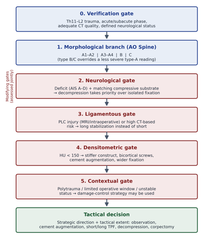
Figure 1 The general sequence of the algorithm: the verification gate and the morphological branch set the baseline tactics, while the neurological, ligamentous, densitometric, and contextual gates modify its scope. The technical level of surgical execution is placed outside the algorithm.
The algorithm's input variables are divided into mandatory ones, without which branch selection is impossible, and modifying ones, which change the scope of intervention or the monitoring regimen. Their algorithmic roles are summarized below:
The tactical extent is described through the construct class and the M1–M4 configurations, whose interpretation depends on whether short or long TPF is meant (Table 5). For corpectomy, the same M1–M4 labels refer to the posterior TPF configuration accompanying the anterior cage, rather than to the cage itself, with cross-linked long constructs (M3–M4) being biomechanically preferred.
|
Construct class |
M1 |
M2 |
M3 |
M4 |
|
Short TPF |
short monocortical screws, no intermediate |
long bicortical screws, no intermediate |
short screws + intermediate screws into the injured vertebra |
long bicortical + intermediate screws |
|
Long TPF |
short monocortical, no cross-links |
long bicortical, no cross-links |
short screws + cross-links |
long bicortical + cross-links |
Table 5 Interpretation of the M1–M4 configurations
Working threshold reference points were defined for each branch (Table 6). In A1–A2, the key branch points are the morpho-functional variant (compensated / subcompensated / decompensated), the risk of kyphosis progression, HU, and pain: compensated injuries are managed predominantly conservatively, whereas decompensated ones require active stabilization. In A3–A4, the algorithm formalizes the choice between indirect and direct decompression, between short and long TPF, and between body preservation and corpectomy by criteria of morphotype, predicted residual stenosis, PLC, deformity, PED, and HU. For type B, long fixation is the baseline, since compromise of the tension structures limits the applicability of short constructs; in B2, the deformational, channel-compressing, and mixed variants must be distinguished by PED and GA thresholds. For type C, the main criterion is the ability to restore the segment's axis and preserve the anterior column.
|
Type / parameter |
Working value |
Tactical interpretation |
Source / status |
|
A1–A2, compensated |
CA < 8.1°; GA < 15.6°; A/P ratio > 0.70 |
Baseline conservative treatment in the absence of other risks |
cluster-derived working threshold |
|
A1–A2, subcompensated |
CA 8.1–12.5°; GA 15.6–23.3°; A/P ratio 0.52–0.70 |
Zone of individual choice; assessment of progression risk and PLC state |
cluster-derived working threshold |
|
A1–A2, decompensated |
CA > 12.5° or GA > 23.3° or A/P ratio < 0.52 |
Active stabilization tactics in the absence of contraindications |
cluster-derived working threshold |
|
A3–A4, short-TPF limit |
CA ≤ 14° and GA ≤ 25°, intact PLC, no need for open decompression |
Short 6-screw TPF permissible |
FEM-supported + clinical working limit |
|
A3–A4, beyond the limit |
CA > 14° or GA > 25°; significant retropulsion; high PLC risk |
Long 8-screw TPF or corpectomy by the vertebral body state |
FEM-supported + clinical working limit |
|
Level-dependent stenosis limits |
Th11–Th12 ≈ 35%; L1 ≈ 45%; L2 ≈ 55% |
Reference for caution regarding decompression (with AIS and predicted residual stenosis) |
residual-stenosis model |
|
B2, by variant |
PED < 3.0 mm / PED ≥ 3.0 mm / PED ≥ 3.0 mm and GA ≥ 27.0° |
Long TPF; decompression by the combination of PED and GA; for the mixed variant — assess the anterior column |
PLC model + ROC reference points |
|
HU |
< 150 / 150–184 / ≥ 185 |
< 150 — reinforce fixation and augmentation; ≥ 185 — standard support in the absence of other risks |
densitometric fixation-risk modifier |
|
Loss of correction / progression |
> 5° from the postoperative or previous value |
Criterion for revising tactics or intensifying monitoring |
clinical safety reference |
Table 6 Working threshold reference points used in the algorithm
Note: The threshold values of CA, GA, and A/P ratio for the A1–A2 morpho-functional variants are a clinically convenient approximation of the cluster boundaries; the final assignment is based on the combination of the three parameters rather than on the isolated exceedance of one of them. CA — Cobb angle; GA — Gardner angle; PED — posterior edge distraction; AED — anterior edge distraction; HU — Hounsfield bone density; TPF — transpedicular fixation.
Branch-specific tactical rules
Compression injuries A1–A2: The main task of this branch is not to miss hidden instability, to separate stable cases for conservative treatment from cases with a high progression risk, and to choose correctly between cement augmentation and posterior stabilization. At the verification step, signs of posterior element injury, facet distraction, widening of the interspinous space, or a high CT-based PLC risk move the case out of the simple A1–A2 branch (MRI is performed or the B2 logic is applied). Assignment to the compensated, subcompensated, or decompensated variant is made by the combination of CA, GA, and A/P ratio: when parameters diverge, priority is given to the most unfavorable one. For A1, the risk of kyphosis progression is assessed by a nomogram at an early follow-up (weeks 2–3) rather than as an urgent test on admission.
The compensated variant is a zone of predominantly conservative treatment (bracing, analgesia, early monitoring); active intervention here is an exception requiring separate justification (persistent pain impeding mobilization, progression beyond 5°). The subcompensated variant is the main zone of tactical uncertainty: the decision is made not by the A1/A2 subtype but by the combination of progression risk, pain profile, HU, expected mobility, and PLC state (from conservative management and cement augmentation to short 4-screw TPF). The decompensated variant, in the absence of contraindications, requires active stabilization - by default short 6-screw TPF with intermediate screws (M3), with reinforcement to M4 and selective augmentation at HU < 150 or high mechanical exposure.
Burst injuries A3–A4: The tactical decision in A3–A4 consists of three interrelated questions: whether decompression is needed, whether short stabilization with preservation of the injured vertebral body is possible, and whether the injured vertebral body has realistic consolidation potential. The algorithm proceeds from the priority of preserving the patient's own vertebral body until this creates a risk of residual stenosis and mechanical insufficiency. A translational or rotational component shifts the case to type C, and a confirmed or high-risk PLC injury shifts it to long stabilization. A kyphotic-deformational variant (predominance of angular deformity) and a compression-retropulsive variant (less deformity with greater PED and a leading role of the posterior wall and canal) are distinguished.
The decision on decompression is made not by the absolute magnitude of stenosis but by the combination of the injury level, AIS, the nature of the compressive substrate, PED, and the predicted residual stenosis after indirect decompression; level-dependent stenosis reference points (≈35% for Th11–Th12, 45% for L1, 55% for L2) are used as a signal of heightened caution. To predict the efficacy of indirect decompression, a residual-stenosis model Θafter = Θbefore − ΔΘ(predicted) is applied. Short 6-screw TPF M4 is permissible only with an intact PLC, no deficit, CA ≤ 14°, GA ≤ 25°, sufficient indirect decompression, and preserved consolidation potential; exceeding these limits, the need for open decompression, or PLC injury shifts to long 8-screw TPF M4. Corpectomy is not a routine consequence of the A4 subtype: it is indicated only for gross multifragmentary body destruction, residual significant stenosis disrupting body support, or the need to remove retropulsed fragments with loss of load-bearing capacity.
Tension-structure injuries (type B): Type B differs from type A by disruption of the tension structures, so even a small angular deformity is not proof of stability, and the baseline principle is long stabilization with additional assessment of the bony component by the logic of the corresponding A subtype. Distractive B1 with a predominantly bony component requires long 8-screw TPF M4 and parallel assessment of the anterior column. B2 is the key subtype, as it reflects a functionally significant injury of the posterior tension complex: short fixation is insufficient even with non-critical deformity, and three variants are distinguished by PED and GA thresholds: a deformational variant (PED < 3.0 mm) requires long TPF M4 with decompression only for clinically significant stenosis; a channel-compressing variant (PED ≥ 3.0 mm, GA < 27.0°) - long TPF M4 with assessment of decompression by AIS, stenosis, and compressive substrate; a mixed variant (PED ≥ 3.0 mm, GA ≥ 27.0°) - long TPF M4 with a high probability of decompression and assessment of the anterior column and the need for corpectomy. The classification thresholds of the variants differ from the ROC reference points of severe neurological deficit (PED ≈ 4.6 mm; GA ≈ 24.0°), which are used specifically for caution regarding decompression. B3 forms via a hyperextension mechanism, often on an ankylosed spine; owing to the limited empirical base, the corresponding recommendations should be regarded as lower-level guidance because of the limited empirical base, with an emphasis on long-lever instability.
Translational-dislocation injuries (type C): Type C is the most unstable owing to the translational component, so the algorithm does not pose the question of whether stabilization is needed but determines whether the segment's axis can be restored by long posterior instrumentation with body preservation, or whether corpectomy with anterior column reconstruction is required. Spatial variants are distinguished by the presence of lateral displacement and rotation. The single-plane variant is the only one in which preservation of the injured vertebral body may be a baseline strategy, but this is confirmed intraoperatively (the axis must be restored and the body must retain consolidation potential); otherwise, the procedure shifts to corpectomy. For the multi-plane and gross-destructive variants, corpectomy is the baseline precisely because of the morphological impossibility of preserving the body rather than for additional correction; at low HU, measures to reduce the subsidence risk are mandatory (augmentation of adjacent endplates, an implant with an enlarged support surface), and for two-level corpectomy the caution threshold is extended to HU < 180–200.
Retrospective decision-level evaluation
43 endpoint events were recorded (Table 7) - 41 predefined structural failures and 2 clinico-deformational ones. Endpoint events were analyzed among the 297 patients with a 12-month follow-up; the generalized adverse clinico-radiological outcome, combining the clinically significant cases of both categories, occurred in 38 patients (the difference from the number of events is explained by the fact that some structural events did not reach the level of a clinically significant adverse outcome and that some patients had more than one event). By decision level, 40 of the 41 structural failures were assigned to the tactical level; no isolated strategic-level failures, in which the initial treatment direction would contradict the basic morphology, were found. Thus, the main zone of potential improvement lies not in the choice between intervention and its refusal but in refining the scope, configuration, and limits of the tactics.
|
Injury type |
n / follow-up |
Structural failure |
Clinico-deformational |
Adverse outcome |
Events mapped to tactical level |
|
A1–A2 |
57 / 53 |
3 (5.7%) |
2 (3.8%) |
5 (9.4%) |
5 |
|
A3–A4 |
100 / 88 |
16 (18.2%) |
0 |
15 (17.0%) |
15 |
|
B |
124 / 117 |
14 (12.0%) |
0 |
11 (9.4%) |
14 |
|
C |
45 / 39 |
8 (20.5%) |
0 |
7 (17.9%) |
8 |
|
Total |
326 / 297 |
41 (13.8%) |
2 (0.7%) |
38 (12.8%) |
42 |
Table 7 Endpoint events and decision-level distribution by injury type
Note: Structural events were assessed among patients with available follow-up. Of the 41 structural failures, 40 were assigned to the tactical level and 1 was non-classifiable; the 2 clinico-deformational failures were also tactical. Cage subsidence (all after corpectomy) accounted for 6 events.
A separate cross-cutting point of tactical uncertainty is corpectomy: in the retrospective analysis, it was associated with the numerically highest observed rate of structural failure among all methods (6 of 23; 26.1%; 95% CI 10.2–48.4%; in all events - interbody cage subsidence; given the small subgroup, the estimate is descriptive) in the absence of an advantage in neurological recovery (AIS improvement in 11/23 (47.8%) versus 111/217 (51.2%) for methods without corpectomy; AIS improvement was assessed only among patients with a baseline deficit [AIS A–D] and paired 12-month documentation). Structural-failure and adverse-outcome rates by treatment method are shown in Table 8. Accordingly, in the algorithm corpectomy is applied only for narrow morphological indications - when body preservation is morphologically impossible - rather than for additional correction.
|
Treatment method |
n |
12-mo follow-up |
Structural failure, n (%) |
Adverse outcome, n (%) |
|
Conservative |
13 |
11 |
0 (0.0) |
2 (18.2) |
|
Cement augmentation |
41 |
37 |
4 (10.8) |
4 (10.8) |
|
Short TPF |
54 |
50 |
12 (24.0) |
12 (24.0) |
|
Long TPF, no decompression |
19 |
17 |
0 (0.0) |
0 (0.0) |
|
Long TPF + decompression |
176 |
159 |
19 (11.9) |
16 (10.1) |
|
Corpectomy |
23 |
23 |
6 (26.1) |
4 (17.4) |
|
Total |
326 |
297 |
41 (13.8) |
38 (12.8) |
Table 8 Structural failure and adverse outcome by treatment method (n = 326; 12-month follow-up n = 297)
Note: Percentages are calculated relative to the number of patients with 12-month follow-up. Methods are pooled across AO Spine types; comparisons are descriptive and unadjusted because treatment groups were not randomized and differed in baseline morphology. Corpectomy includes single- and two-body procedures.
Typical problem scenarios and the corresponding algorithm recommendations that illustrate the tactical level of potential improvement:
For practical use, the key branch points are summarized in a short checklist (a “yes” answer to each question sets the corresponding action):
Software implementation
To increase the reproducibility and practical applicability of the algorithm, a software implementation was created in the form of an interactive web-based clinical decision-support interface (Figure 2). The interface is not a new classification system or an independent protocol; its purpose is to reproduce the algorithm's logic step by step with transparent display of the active if–then rules. The implementation is modular, with each module corresponding to an analytical stage: primary AO Spine classification, entry of neurological status, morpho-functional refinement of the injury variant, CT-based assessment of PLC risk when the PLC state is uncertain, selection of the tactical branch (conservative management, cement augmentation, short or long TPF, decompression, corpectomy), technical refinement of the stabilization configuration (construct length, screw type, intermediate screws or cross-links, augmentation by HU), and determination of biomechanically unfavorable postoperative loading patterns.
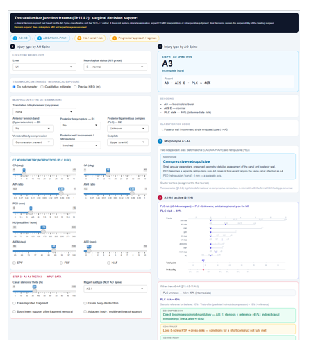
Figure 2 Screenshot of the software implementation of the individualized treatment-planning algorithm as a web-based clinical decision-support interface.
For each tactical branch, the interface displays not only the final recommendation but also the list of activated rules and factors that led to it, implementing the principle of decision transparency and avoiding “black-box” logic. The main purpose of the implementation is to standardize the sequence of clinical-morphological analysis, increase the transparency of decision-making, and reduce the risk of missing critical features that change treatment tactics. The interface remains a decision-support tool and does not replace clinical examination, MRI, expert CT assessment, or the surgeon's intraoperative decision. A working version is available online at https://alexeyns.shinyapps.io/algo_en/.
The main practical result of the evaluation is that the algorithm mainly refines the tactical rather than the strategic level of a decision: short fixation is replaced by long, stabilization is supplemented by decompression, preservation of the injured vertebral body is replaced by corpectomy under appropriate indications, and standard monitoring is intensified in patients with low HU or high mechanical exposure. The predominance of tactical-level failures (40 of 41) and the absence of isolated strategic-level failures are consistent with the fact that current classifications fairly reliably determine the indication for intervention but provide little formalization of its scope.2,5 The choice between short and long fixation and the factors of failure of short-segment constructs remain subjects of active study.20,21 The separation of strategic, tactical, and technical levels has methodological value because it prevents adverse outcomes from being interpreted as a single homogeneous category. In this cohort, most structural failures mapped to tactical branch points, suggesting that the main modifiable zone is the extent and configuration of treatment rather than the initial decision to intervene.
The relatively high observed frequency of subsidence after corpectomy in the absence of a neurological advantage underscores that expanding the scope of intervention is not an end in itself: the decision on anterior column reconstruction should be based on the morphological impossibility of preserving the body rather than on a desire for additional correction. At the same time, these data should be interpreted with allowance for probable confounding by indication: corpectomy was performed predominantly for the most severe morphological scenarios - gross body destruction, lower HU, and greater loss of anterior support - so the higher subsidence rate cannot be unconditionally attributed to the method itself, as it may reflect a more severe baseline profile of these patients. This is consistent with biomechanical considerations of the safety margin of reconstructive constructs and with the guidance to apply the most invasive solutions only for clear morphological indications.
The proposed algorithm does not compete with AO Spine or TLICS but complements them: it uses the type and severity of the injury as input data and then integrates the morpho-functional variant, the canal and PLC state, HU, and the biomechanical limits of constructs into a sequence of tactical choice. The separation of the strategic, tactical, and technical levels makes it possible to analyze separately the sources of adverse outcomes and to document transparently the grounds for a specific decision - why conservative management, cement augmentation, short or long TPF, decompression, or corpectomy was chosen.
In practical terms, the algorithm’s role begins after the injury type and the general indication are established: it helps determine whether short fixation is sufficient, whether decompression should be added, whether low HU requires reinforcement of the construct, and whether corpectomy is morphologically unavoidable. The study has limitations. The algorithm was formed on a single-center retrospective-prospective cohort; individual prognostic models have only internal validation, so the quantitative thresholds should be interpreted as working decision-support limits rather than absolute indications for a particular operation. In particular, the values of CA, GA, PED, and HU should be regarded not as automatic commands for a specific intervention but as reference points for reducing uncertainty, whereas the final decision integrates the combination of features and the clinical context. The retrospective evaluation demonstrates a logical correspondence between the structure of decision-level failures and the algorithm's branch points but does not prove that the algorithm would have prevented adverse outcomes. The decision-level classification was performed retrospectively and, despite independent expert assessment and consensus agreement, does not fully exclude subjectivity; prospective independent assessment would strengthen this result. The algorithm does not model situations of an absolute priority of life-saving measures (damage control) and does not cover the technical level of surgical execution, failures of which require a separate analysis of surgical quality. The long treatment period (2011–2025) may have introduced temporal heterogeneity in surgical technique, implant selection, imaging availability, and postoperative management. The study did not directly quantify inter-surgeon variability in decision-making. Overall, this evaluation should be interpreted as a structured audit of decision logic rather than as evidence of clinical effectiveness.
The algorithm should be regarded as a tool for risk stratification and decision support that makes the sequence of tactical choice transparent and reproducible and reduces the variability of approaches. Further implementation requires prospective verification of its effect on the rates of structural failure, reoperation, loss of correction, and functional outcomes, as well as external validation of the thresholds on independent cohorts.
An individualized treatment-planning algorithm for traumatic thoracolumbar junction injuries was developed by integrating AO Spine morphology, morpho-functional injury variants, neurological status, canal compromise, PLC-risk assessment, HU, mechanical exposure, and biomechanical construct limits into a sequential if–then framework. In retrospective decision-level evaluation, most observed structural failures mapped to tactical branch points rather than to isolated strategic-level failures. Corpectomy was associated with a high observed rate of cage subsidence and should be reserved for narrow morphological indications. The algorithm should be regarded as a transparent decision-support framework rather than a rigid protocol, and its clinical effectiveness requires prospective external validation.
The study was performed as part of a planned research project (state registration number 0119U000110); no targeted external funding was involved.
None.
The authors declare that there are no conflicts of interest.
The data supporting the findings of this study are available from the corresponding author upon reasonable request, subject to restrictions related to patient confidentiality.

©2026 Nekhlopochyns, et al. This is an open access article distributed under the terms of the, which permits unrestricted use, distribution, and build upon your work non-commercially.
