Journal of
eISSN: 2373-4426


Case Report Volume 16 Issue 1
 Maria Cristina Quevedo Villaseñor
Independent researcher, Mexico
Correspondence: María José Salazar Torres, Independent researcher, Mexico, Tel +52 55 27 29 71 31
Received: February 24, 2026 | Published: March 10, 2026
Citation: Torres MJS, Villaseñor MCQ. Neonatal arterial ischemic stroke involving the middle cerebral artery territory: a case report and literature review. J Pediatr Neonatal Care. 2026;16(1):20-24. DOI: 10.15406/jpnc.2026.16.00613
A female newborn, born at 38+2 weeks of gestation with the following maternal history: 33-year-old mother, first pregnancy, diagnosed with inappropriate sinus tachycardia in 2023, treated with metoprolol. Maternal vaccination schedule was complete according to age. Maternal history negative for tobacco, alcohol, or illicit drug use.
Perinatal history: normal pregnancy with prenatal care from the beginning of gestation, 12 obstetric ultrasounds, the last at 38 weeks of gestation reporting a single female pregnancy with an approximate weight of 2750 grams, placenta and umbilical cord without abnormalities. Amniotic fluid with decreased total volume. First and second trimester markers not performed. Screening for gestational diabetes negative. Vaginal cultures negative. Baby was born via vaginal delivery with clear amniotic fluid, she was reactive and good tone, with Apgar 9/9, Silverman Anderson 0. Immediate skin-to-skin contact began, where initial neonatal resuscitation steps were completed, to which she responded adequately. Early maternal attachment happened for 30 minutes. No abnormalities on physical examination. She underwent a transition period without complications, remaining in joint accommodation with her parents with breastfeeding and supplemented with starter formula, reporting adequate urine output and spontaneous bowel movements. Cardiac and hearing screening tests were performed and reported normal.
During her stay in the physiological care nursery, in the first hours of life she presented weak sucking and fatigue, for which suction therapy was indicated once a day. At 36 hours of life, clonic movements were reported in the left hand, followed by the ipsilateral and then contralateral hemibody, lasting less than one minute, and the latter associated with cyanosis and desaturation up to 60%, therefore she was admitted to the neonatal intensive care unit for proper management.
Upon admission, vital signs were within normal parameters, with no need for supplemental oxygen, no evidence of respiratory distress, adequate respiratory pattern, hemodynamically stable, and no clinical gastrointestinal abnormalities. Physical examination revealed normotensive fontanelles, no cardiopulmonary compromise, and primitive reflexes present (Moro, rooting, suction, palmar and plantar grasp), well developed extremities, adequate tone and strength. No persistent neurological focus. Monitoring was initiated with integrated amplitude electroencephalography (aEEG), presenting during the first hours an event of desaturation (80%) accompanied by brief eye movements (<6 seconds) and intermittent sucking, without electroencephalographic correlation. A transfontanellar ultrasound was performed, reporting a structurally intact brain, no edema, hemorrhage, or areas of ischemia, with a resistance index of 0.78. A one-hour video electroencephalogram (vEEG) documented four electrographic events in the right frontotemporal region and interictal activity with sharp waves and slow angular waves in the same region. Phenytoin infusion at 20 milligrams/kilogram/day was administrated. Additional studies: Venous blood gas analysis with acid-base balance, lactate 3.7 mmol/L, and anion gap 16. Complete blood count without signs of infection, negative acute phase reactants, blood chemistry with phosphate: 5.65 mg/dL, potassium: 5.53 mEq/L, lactate dehydrogenase (LDH) of 677 U/L, total bilirubin: 9.58 mg/dL, no other biochemical alterations.
She was later evaluated by pediatric neurology, which found eutrophic extremities, slightly decreased tone, preserved strength, deep tendon reflexes 2+/4+, bilateral plantar extensor response, no clonus. Maintenance dose of phenytoin at 8 milligrams/kilogram/day was continued and an MRI with gadolinium under sedation was requested.
At 72 hours of life, a brain MRI was performed, which showed acute bilateral ischemic stroke in the middle cerebral artery territory, more extensive in the right hemisphere, with no evidence of hemorrhage. Magnetic resonance angiography showed no obvious obstructions (Figures 1–5).
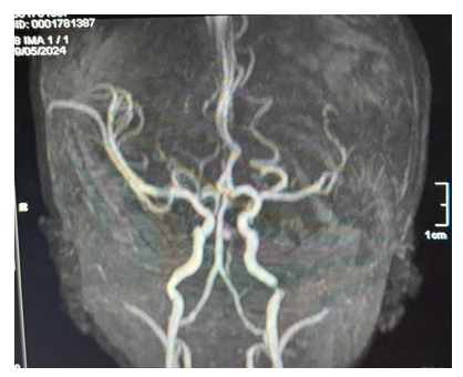
Figure 1 Magnetic resonance angiography.
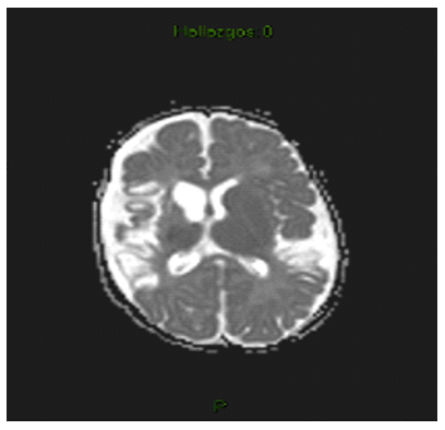
Figure 2 Axial MRI scan with hyperintense areas.
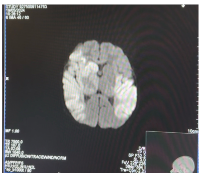
Figure 3 Axial section of both cerebral hemispheres.
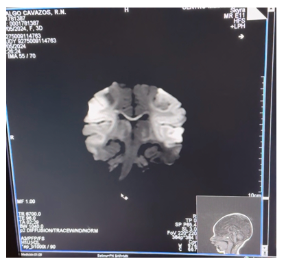
Figure 4 Coronal section of cerebral hemispheress.
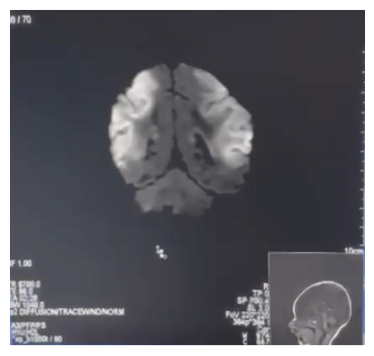
Figure 5 Coronal section of the frontotemporal region.
Given the finding of bilateral middle cerebral artery infarction, the clinical approach was complemented with an echocardiogram, which reported normal structural anatomy, minimal patent foramen ovale (<1 mm) with left-to-right shunt, no structural congenital heart disease, preserved biventricular function, and no evidence of pulmonary hypertension. Carotid ultrasound showed no abnormalities.
A consultation with pediatric hematology was requested, initiating an etiological approach aimed at neonatal thrombophilia. Initial laboratory tests: D-dimer: 1650 ng/mL, fibrinogen: 405 mg/dL, homocysteine: 5.9 µmol/L, normal coagulation times, complete blood count without thrombocytosis or thrombocytopenia.
Placental study showed no necrosis or significant changes. Anticoagulation was started with enoxaparin 1.5 milligrams/kilogram every 12 hours empirically given the bilateral extent of the ischemic event, with subsequent determination of anti-Xa levels within the therapeutic range.
The thrombophilia profile included: Antithrombin III: 54% (within the expected range for neonatal age), lupus anticoagulant: negative, antinuclear antibodies: negative, normal anticardiolipins, TORCH profile: negative. Factor V Leiden mutation, prothrombin mutation, methylenetetrahydrofolate reductase (MTHFR) mutation with normal homozygous result.
At 5 days of age, she was evaluated by rehabilitation medicine, which found: tendency toward axial and appendicular hypotonia, absent rooting reflex, weak sucking with fatigue (36 sucks/minute, brief pauses), incomplete Moro reflex, popliteal angle 120°, adductors 140°, dorsiflexion -20°, weak plantar grasp, no clonus. A neonatal neurological therapy program was started twice a day with the following objectives: tone management, early kinesitherapy, multiple sensory stimulation, and proprioceptive stimulation.
In the absence of documented prothrombotic factors and clinical stability, it was later decided to suspend anticoagulation on the seventh day of life, considering that the risk of bleeding outweighed the potential benefit. During the following days, she remained hemodynamically stable, and the transition from intravenous to oral phenytoin was made without recurrence of seizures. A follow-up EEG video showed abnormal grade III activity with sharp and slow angular waves in the right frontotemporal region, without electroclinical seizures. Serum phenytoin levels were documented within the therapeutic range.
On the eighth day of life, she remained stable, no need of oxygen, breastfed with adequate suction-swallowing, weighing 2635 grams, neurologically stable, and therefore it was decided to discharge her, with follow-up by neonatology, neuropediatrics, hematology, and rehabilitation.
Magnetic resonance imaging (MRI) control after discharge, at 4 months of age, identified areas of malacia and multilobar corticosubcortical gliosis, encephalomalacia, and corticosubcortical gliosis of the right fronto-parieto-temporal and left frontoparietal regions, as sequelae of a vascular event in the territory of the bilateral ACM. Angiography showed filling of both middle cerebral arteries. There is a fetal variant of the left posterior cerebral artery. Both internal carotid arteries and the vertebrobasilar system are adequately filled.
She is currently under close neurodevelopmental surveillance. At 21 months of age, the patient's overall neurodevelopment is appropriate for her age, with no clinical evidence of focal motor sequelae, epilepsy, or cognitive disorder, and no evident. Neurological examinations by pediatrics and neurology to date are normal for her age.
Perinatal ischemic infarction is defined as an event that interrupts cerebral blood flow due to an arterial or venous thrombus or embolus, occurring between the 20th week of gestation and the first 28 days of life, with an estimated prevalence of 1 per 1,100–2,500 live births according to the literature consulted. It represents one of the main causes of acquired focal brain injury in newborns.1–5
Because this condition remains underdiagnosed and its exact pathophysiology is not fully understood, there are few preventive measures and even fewer therapeutic options.3
Perinatal arterial ischemic infarction (PAIS) is the most common type of perinatal ischemic infarction and usually occurs in the first days of life with seizures in the first 12-72 hours (70-90% of patients) or with more nonspecific findings such as encephalopathy, absence of tone, or sucking disorders.1,2
Although it was previously considered a relatively rare condition, with the widespread use of advanced neuroimaging techniques, it is now known that many cases that were previously classified as idiopathic or congenital cerebral palsy are due to perinatal infarction.2,6
The most frequently affected vascular territory is the middle cerebral artery (MCA), with unilateral predominance (the left side being the most frequent),2 and it has recently been proven that infarcts involving large areas of the MCA, including complete hemispheric infarcts, are usually associated with a higher risk of significant sequelae, as well as some type of long-term disability.7
Infarcts involving the entire MCA territory are a rare clinical variant but are highly relevant due to their association with significant cerebral edema and an increased risk of neurological sequelae.7
The case mentioned above corresponds to an extensive bilateral infarction with a good neurological prognosis, which is an uncommon presentation.
Etiology
The etiology of PAIS is multifactorial, and its underlying mechanisms remain incompletely understood,1 but maternal, obstetric, anatomical, and genetic causes have been described.8 Likewise, the presence of maternal or neonatal systemic inflammation could intensify brain damage.6,9 Unlike stroke in adults, in newborns, it can rarely be attributed to a single identifiable cause. Several studies have shown that the vast majority of cases remain without a single defined etiology.10 Even so, in most of the studies that have been conducted, at least one risk factor is present in the majority of cases (82%).10 That being said, one of the most widely accepted theories today suggests that PAIS is the result of the interaction between inflammation, thrombosis, and cerebral vascular vulnerability. This theory would explain why many patients have multiple risk factors rather than a single obvious cause.2,9
Maternal factors: Several maternal factors have been associated with an increased risk of perinatal infarction, particularly those related to systemic inflammation and coagulation disorders. These include infections during pregnancy, hypertensive disorders, gestational diabetes, and autoimmune conditions,10 as well as primiparous mothers, emergency cesarean section, and chorioamnionitis.6,8,11 It is believed that the conditions mentioned above can induce endothelial activation and changes in the placental coagulation cascade, promoting the formation of emboli that subsequently reach the fetal cerebral circulation.6,12
Although in the case mentioned in this article, pathology determined that the placenta did not present any pathological findings, several studies have frequently identified lesions compatible with poor fetal vascular perfusion, placental thrombosis, and chronic inflammation in neonates with PAIS. All these findings support the hypothesis of embolism originating in the placenta, especially in cases where the newborn has no apparent cardiac or vascular abnormalities.4,13
Neonatal factors: Risk factors in the newborn include transient prothrombotic states, polycythemia, congenital heart disease, and hemodynamic alterations during the peripartum period, as well as the use of forceps, neonatal hypocalcemia, prolonged or instrumental labor, perinatal asphyxia, and an Apgar score below 7.6,8,11,12
Pathophysiology
Although it is known that PAIS is secondary to arterial obstruction, the cause of this occlusion has not yet been identified. Unlike ischemic stroke in adults, where the pathophysiology is well identified and usually secondary to atherosclerosis or hypertension, the etiology of PAIS is related to factors that occur before or during labor, and although it is not yet well understood, the hypotheses most frequently mentioned in the literature include placental thromboembolism, maternal-fetal inflammation, or an intracranial or carotid injury during delivery.4,11
The neonatal brain has structural and metabolic characteristics that condition a particular response to ischemic damage. Compared to the adult brain, it has greater neuronal plasticity; however, it is also more susceptible to cellular injury mechanisms. The immaturity of antioxidant systems and the high density of excitatory receptors mean that even brief periods of hypoperfusion can trigger significant neuronal damage.4,9,11
Clinical manifestations and initial presentation
Neonatal seizures are the most common manifestation of neonatal arterial infarction and usually occur within the first 72 hours of life. Unlike epileptic seizures in later stages, they are usually focal and may manifest as subtle clonic movements, ocular deviation, or brief motor episodes that are difficult to recognize clinically, which may lead to underrecognition of seizure activity. Other signs include hypotonia, sucking difficulties, apnea, motor asymmetry, and altered consciousness.5,13,14
However, some newborns remain clinically stable during the immediate neonatal period, and the diagnosis is made later when abnormalities or delays in neurological development are detected.1,9,11
Diagnosis
The diagnosis is clinical and supplemented by neuroimaging and a high index of clinical suspicion should be maintained in all newborns who, in the presence of any risk factor, present with any of the neurological abnormalities described above. Magnetic resonance imaging (MRI), especially when performed 2 to 4 days after the injury, is considered the gold standard due to its high sensitivity (about 91%) in detecting even very small infarcts, in addition to being a non-invasive procedure that does not use radiation. In addition to this, it is important to mention that MRI can detect the affected vessels without the need for contrast, which can identify the cause of the infarction and, as mentioned previously, predict the prognosis.15,16
However, the clinical-radiological correlation in neonates is less predictable than in adults due to the plasticity of the developing brain. This variability explains why apparently extensive lesions may be associated with favorable functional outcomes in some patients, as is the case presented in this article.7,8
Transfontanellar ultrasound remains a useful tool as an initial study, although its sensitivity is limited (30.5%) for detecting small cortical lesions or early infarcts, in addition to being operator-dependent. However, as it is a non-invasive study that can be performed in real time at the patient's bedside, it continues to be widely used as a preliminary initial study.11
Prognosis
The long-term prognosis is difficult to predict, as the results obtained in the reviewed literature were highly variable, depending on the type of infarction, the type of study, the length of follow-up of patients with some type of neonatal infarction, the time of onset, and the type and duration of rehabilitation and physical therapy, among other factors.3 Another important variable to consider in the long-term prognosis is brain neuroplasticity, which plays a decisive role, as well as early rehabilitation.
In general terms, PAIS is one of the main causes of hemiplegic cerebral palsy, and it is estimated that about 60% of those affected will have some type of motor deficit and about 50% will have some behavioral alteration, although these do not always manifest themselves within the first years of life.1 In addition to motor impairments, patients may experience cognitive difficulties, language disorders, and epilepsy, which may develop months or years after the initial event.5,10 A review of the literature reports a mortality rate of 3%.9
Approach and management
In most cases, the approach to a newborn with an infarction is not limited to imaging studies to confirm the diagnosis; additional diagnostic evaluations are also performed to determine the etiology of the event. Although placental histopathology can be a key factor in identifying the etiology of the infarction, there are few studies in which any abnormalities were found.8 Other complementary studies include a cardiological evaluation with echocardiogram, as well as genetic or hematological counseling to look for thrombophilic disorders.8 Of all the studies reviewed, only one retrospective study of 55 newborns was found in which 70% of patients had placental histopathological abnormalities and 87% had some form of heart disease that could explain the etiology of the neonatal infarction.8
The management of patients with PAIS depends largely on their clinical condition. In addition to symptomatic management, in patients who have seizures, a continuous integrated amplitude electroencephalogram can be used, especially to assess the efficacy of anticonvulsant drugs. Routine anticoagulation is not recommended.16
Although PAIS is underdiagnosed due to the clinical challenge it represents, its incidence is increasingly recognized as clinically significant and it has a significant impact on neurodevelopment and long-term quality of life, as well as being a potentially disabling condition.
In the presence of any neurological abnormality in a newborn without apparent cause, it is vitally important to have a high index of suspicion of neonatal arterial infarction as the underlying cause, as early diagnosis and multidisciplinary intervention could favorably influence the neurological prognosis.
None.
None.
The authors declare that they have no competing interests.

©2026 Torres, et al. This is an open access article distributed under the terms of the, which permits unrestricted use, distribution, and build upon your work non-commercially.