Journal of
eISSN: 2373-4426


Case Report Volume 15 Issue 3
1Pediatric Surgeon, Hospital Infantil Universitario de San José, Colombia
2Pediatric Surgery Resident, Fundación Universitaria de Ciencias de la Salud – FUCS, Colombia
3Pediatric Surgeon, Airway and Thoracic Pediatric Surgery, Hospital Infantil Universitario de San José, Colombia
4Pediatrician Neonatologist, Hospital Infantil Universitario de San José, Colombia
5Pediatrics Resident, Fundación Universitaria de Ciencias de la Salud – FUCS, Colombia
6Medical Student, Fundación Universitaria de Ciencias de la Salud – FUCS, Colombia
7General Physician, Fundación Universitaria de Ciencias de la Salud – FUCS, Colombia
Correspondence: Shary Acosta Suárez, Pediatric Surgeon, Hospital Infantil Universitario de San José, Cra. 52 # 67a - 71. Fundación Universitaria de Ciencias de la Salud – FUCS, Bogotá, Colombia, Tel (+57) 3214772200
Received: August 22, 2025 | Published: September 9, 2025
Citation: Acosta S, Mendoza DI, Romero DP, et al. Intrathoracic H type fistula, a diagnostic challenge: case report . J Pediatr Neonatal Care. 2025;15(3):142-144. DOI: 10.15406/jpnc.2025.15.00599
Background: Isolated H-type tracheoesophageal fistula (TEF) is a rare congenital anomaly, accounting for less than 5% of tracheoesophageal malformations. It typically presents with choking, coughing, and cyanosis during feeding. However, diagnosis is often delayed due to its non-specific presentation and the intermittent nature of symptoms. This case highlights the diagnostic challenges and surgical management of an intrathoracic H-type TEF located below T4, an even rarer finding.
Methods: We report the case of a full-term newborn who developed respiratory distress and cyanosis shortly after birth. Initial imaging, including an esophagogram, was inconclusive. Due to persistent symptoms, bronchoscopy was performed, revealing an H-type tracheoesophageal fistula. Surgical correction was performed via thoracotomy with direct fistula resection, tracheoplasty, and esophageal repair.
Results: The patient was successfully extubated postoperatively but developed airway edema and croup, requiring reintubation. A follow-up esophagogram confirmed intact surgical repair with no residual fistula, malacia, or stenosis. Oral feeding was initiated on postoperative day 7, and the patient was discharged with full oral tolerance and supplemental low-flow oxygen.
Conclusion: Intrathoracic H-type TEF remains a diagnostic challenge due to its atypical location and variable presentation. This case underscores the importance of bronchoscopy in diagnosis and highlights surgical strategies to optimize outcomes. Early recognition and multidisciplinary management are crucial to prevent complications and ensure long-term airway and esophageal function.
Keywords: tracheoesophageal fistula, congenital anomalies, bronchoscopy, neonatal surgery, respiratory distress, case report
Congenital tracheoesophageal fistulas are rare conditions, H-type, N-type or isolated fistula was described in 1873, it is defined as an isolated tracheoesophageal fistula without atresia, it connects the posterior wall of the trachea to the esophagus. It has better outcomes than other fistulas.1 It presents with choking during feeding, cough, cyanosis but its diagnosis is difficult and it usually is delayed until recurrent respiratory infections are presented.2,3 Given the rarity of this entity there are few cases in literature, an incidence less than 6% is reported.2 They represent 4-5% of all tracheoesophageal malformations.3
A newborn born as a result of vaginal delivery at term with spontaneous neonatal adaptation, adequate weight and height at birth, normal prenatal infectious and ultrasound screening. At 4 hours of life presented with cyanosis and respiratory distress after breastfeeding intake, requiring admission to the NICU (neonatal intensive care unit) for low-flow oxygen supply and monitoring. Enteral tube feeding was tolerated so oral suction feeding was scaled, but again, presented severe respiratory distress, ventilatory failure and orotracheal intubation was indicated, pneumonia was suspected. After that, failure to conventional ventilation was presented requiring VAFO (high frequency oscillatory ventilation) and severe pulmonary hypertension was documented in echocardiography ruling out congenital heart disease, was managed with nitric oxide, classified as responder and withdrawal was achieved 48 hours after its initiation.
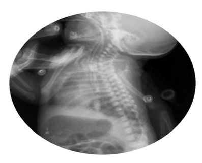
Abdominal radiographs showed persistent severe distension of the gastric chamber despite high caliber tube drainage and evidence on one occasion of bubbling of the same so tracheoesophageal fistula was suspected. Barium esophagogram was performed with evidence of complete passage into esophagus, stomach and pylorus, without a fistulous tract (Figure 1). However, due to persistent symptoms, bronchoscopy was performed to rule out congenital malformation of the airway, H-type tracheoesophageal fistula of 3 mm in diameter located 5 rings above the carina was visualized, nevertheless, endoscopic or surgical correction at this time was not possible due to hemodynamic instability (Figure 2).

Figure 1 Barium contrast esophagogram with no evidence of fistulas or extrinsic compressions. Normal.
Surgical correction was decided, given the location of the fistula, a thoracotomy surgical approach was performed. Initially, the esophagus and trachea were identified, the esophagus was surrounded with vessel loops exposing the fistula, these were placed proximal and distal to the fistula delimiting it completely. (Figure 3). Tracheal plasty was performed on the posterior wall with continuous suture of PDS 5/0, hydrostatic test was realized without evidence of leaks, after which lateral tracheopexy was performed with 2 stitches of PDS 5/0 fixed to the thoracic spine. Finally, closure of the esophageal defect was performed in 2 planes: the first plane with continuous suture of PDS 5/0 and the second with separate invaginating stitches of PDS 5/0 and an orogastric tube was advanced, pneumatic test was performed without evidence of leaks. At 24 hours postoperatively, meets gasometric criteria, radiological and ventilatory parameter criteria for extubation, which was performed. Postoperative bronchoscopy and esophagogram were performed prior to the initiation of the oral route, reported an intact suture without refistulization, confirming the success of the surgical repair and the stability of the airway. The patient was discharged tolerating the oral route by suction and low flow oxygen supply.
Isolated tracheoesophageal fistula is rare, representing less than 5% of all congenital tracheoesophageal malformations, it is classified as E or 5 in the classification of esophageal atresia.1–3 A preponderance of males has been reported with a 2:1 ratio.4,5 More than 70% of the reported fistulas are located above T2,6 in our clinical case it was located intrathoracic below T4 which confers an even more rare finding.
Clinical presentation is variable, coughing, cyanosis, or sudden choking may be evident with feeding, this 3 symptoms are described as a the classic triad. It may also present with recurrent respiratory infections, hypersecretion and recurrent pneumonia. Given the variability of symptoms, there could be patients with very mild symptoms and patients with severe respiratory deterioration requiring mechanical ventilation.1,3,6,7,8
Diagnosis is difficult and often delayed,4,9 definitive diagnosis is made by contrast studies, specially prone tube esophagogram8 or bronchoscopy, these studies define the surgical approach and whether associated tracheal anomalies are found.1,7,10 Bronchoscopy was key in our patient to establish the diagnosis.
Surgical management is imperative to resolve the fistula, endoscopic techniques with sealants have been described,11 but the open approach is more frequent. Cervical approaches are done in fistula locations above T2, in fistulas located T3 or above are done via thoracotomy or thoracoscopy. There is discussion between simple ligation and total fistula resection, other authors also recommend interposition of material between the esophagus and trachea to avoid refistulization.1,4 In our clinical case we decided to resect the fistula, reconstruct the esophagus and tracheal wall, and posterior tracheopexy to avoid recurrence.
Complications include stenosis, anastomotic leak and recurrence, in a long-term gastroesophageal reflux has been reported, which is why follow-up is key in this type of pathology.7,9
Intrathoracic H-type TEF remains a diagnostic challenge due to its atypical location and variable presentation. This case underscores the importance of bronchoscopy in diagnosis and highlights surgical strategies to optimize outcomes. Early recognition and multidisciplinary management are crucial to prevent complications and ensure long-term airway and esophageal function.
None.
Declarations of interest: none.
The authors did not receive support from any organization for the submitted work.
This case report was presented as a poster at the “XXVI Congreso Colombiano de Cirugía Pediátrica”. Cartagena, Colombia April 2th, 2025.
The authors did not receive any financial support for the submitted work.
The authors declare that there is no conflicts of interest.

©2025 Acosta, et al. This is an open access article distributed under the terms of the, which permits unrestricted use, distribution, and build upon your work non-commercially.
 International Childhood Cancer Day is observed on 15 February 2026 to raise awareness about childhood cancers and to highlight the medical, developmental, and supportive care needs of affected children and their families. This day emphasizes the importance of early diagnosis, pediatric care, and continued research to improve survival and quality of life in children with cancer.
Researchers and healthcare professionals are encouraged to submit their original research articles, reviews, and clinical studies related to pediatric oncology, neonatal care, and child health. Manuscripts submitted on the occasion of International Childhood Cancer Day will be eligible for a special publication discount of 30–40% in the Journal of Pediatrics & Neonatal Care (JPNC).
.