Journal of
eISSN: 2373-6410

Introduction: Pure arterial malformations (PAMs) are rare intracranial vascular anomalies characterized by dilated, tortuous, and overlapping arteries without an associated venous component. Although their etiology remains uncertain, most reported cases suggest a benign natural history.
Case report: We describe a 20-year-old woman who presented with severe headaches, photophobia, and scintillating scotomas. Brain imaging performed during childhood had already revealed dilated and redundant arterial segments in the right middle cerebral artery (M1) and posterior cerebral artery (P1/P2), along with a persistent trigeminal artery. Thirteen years later, new imaging - including CT, MRI, and digital subtraction angiography with 3D reconstruction-showed stability of the lesion, absence of aneurysms, arteriovenous shunting, or any ischemic or hemorrhagic changes. The clinical symptoms were attributed to migraine with aura, and conservative management was maintained.
Discussion: PAMs predominantly affect young women and are typically detected incidentally. The middle cerebral artery and posterior circulation are among the most frequent sites. Accurate diagnosis relies on high-resolution angiography to distinguish PAMs from other vascular pathologies, such as arteriovenous malformations or dolichoectasia. Despite their uncertain origin-whether congenital or acquired-available evidence indicates a stable, non-progressive course in most cases.
Conclusion: This report reinforces the benign nature of PAMs and supports conservative management with periodic imaging follow-up as the preferred approach.
Keywords: Pure arterial malformation, cerebral arterial malformation, middle cerebral artery arteriopathy, intracranial vasculopathy, intracranial dilatative arteriopathy, cerebral vascular malformation
PAM, pure arterial malformation; MRI, Magnetic Ressonance Imaging; CT, Computed Tomography; DSA, digital subtraction angiography; PCoA, posterior communicating artery; MCA, middle cerebral artery; PCA, posterior cerebral artery
Pure arterial malformations (PAMs) were first described by McLaughlin et al. in 20131 as dilated, tortuous, and overlapping arteries with a redundant, coil-like appearance. With an estimated low incidence, PAMs are considered rare conditions, reported only in a few individual cases and small case series in the literature. Although some reports describe hemorrhagic complications,2 most of the literature indicates a benign course over the years, suggesting that conservative management is appropriate.3 Here, we report a case of pure arterial malformation that remained stable over 13 years of follow- up.
A 20-year-old woman was admitted to the emergency department of our hospital complaining of intense, limiting, and refractory headaches. The pain had started 5 days prior to hospitalization, occurring daily, and was associated with photophobia, nausea, and scintillating scotomas. She denied fever, seizures, or any focal neurological deficit. Her past medical history was unremarkable for trauma, hypertension, or smoking. At the age of 7, she had undergone brain imaging in another facility due to repetitive hand movement episodes, later diagnosed as a nervous tic. Brain Magnetic Ressonance Imaging (MRI) and digital subtraction angiography (DSA) at that time revealed dilated, tortuous, and redundant right M1 and P1/P2 segments, as well as a right persistent trigeminal artery. The patient was then managed conservatively with observation and recommendations for follow-up MR angiography, which she pursued irregularly every few years.
After hospitalization, the patient’s neurological examination, cerebrospinal fluid analysis, and blood samples were unremarkable. New brain imaging was performed. Computed Tomography (CT) scan and CT angiography showed focal calcification in the right M1 arterial wall (Figure 1). Additionally, brain MRI showed no evidence of intramural hematoma in the arterial walls, nor signs of acute or chronic ischemia, or recent or remote hemorrhage. The abnormal vessels did not appear to have changed over time, and no cortical dysplasia was noted (Figure 2). DSA with 3D reconstruction was requested to better evaluate the anatomy of this vascular anomaly, potential changes in its size or angioarchitecture, and any high-risk features for future bleeding. This study confirmed the absence of aneurysms, nidus, or early venous drainage related to the malformation, as well as the hemodynamic stability of the lesion (Figure 3).
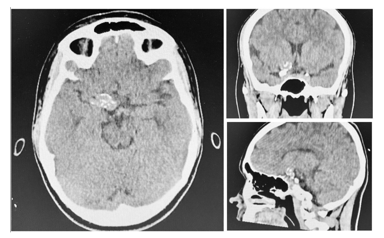
Figure 1 Axial, coronal and sagital CT scans showing focal calcification in right M1 arterial wall.
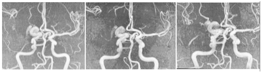
Figure 2 Follow-up brain MRI images in 2016, 2018 and 2025 showing global stability of the arterial malformation.
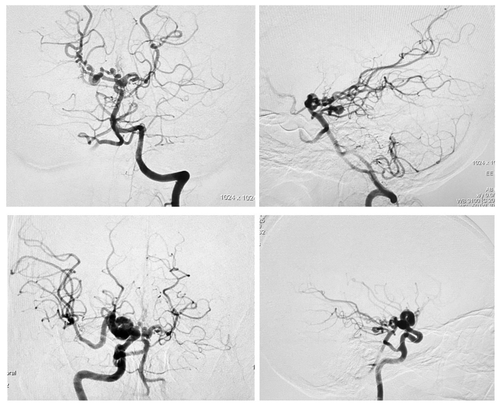
Figure 3 AP and lateral angiograms after injection of the left vertebral artery (upper row) and right internal carotid artery confirming the absence of aneurisms, nidus or early venous drainage related to the malformation and the hemodinamic stability of the lesion.
Given the circumstances, the symptoms were interpreted as migraine with aura, worsened by analgesic overuse, and unrelated to the arterial anomaly. No specific treatment was recommended for the arterial
malformation. The patient was discharged after adequate pain control.
Due to their rarity, the pathophysiology, natural history, and optimal management of PAMs are not well established. According to published cases, this condition exhibits a female predominance, with a median age of 25 years. Patients are generally asymptomatic, and the diagnosis is incidental in over 85% of cases. PAMs can be found in any intracranial artery.2–5 The most common locations are the supraclinoid segment of the internal carotid artery, the posterior communicating artery (PCoA), the anterior cerebral artery, the posterior cerebral artery (PCA) and the M1 segment of the middle cerebral artery (MCA).3,6 Lesions involving the PCoA–PCA or MCA were more likely to present with local aneurysms or calcification, as seen in the case reported here.5
No precipitating risk factor has been definitively associated with this condition, and a history of ischemic or hemorrhagic stroke is unusual, although there is still a lack of long-term follow-up data.3
The etiology remains uncertain. One hypothesis suggests that an acquired cause, such as a viral infection or a somatic mutation later in life, could affect a particularly vulnerable arterial segment. Another proposes that a congenital abnormality or early insult may lead to arterial dysplasia. The congenital theory is supported by Sorenson et al.7 and Brinjikji et al.,3 who reported adipose tissue interspersed within the affected artery.
Often confused with other vascular abnormalities, the diagnostic accuracy of PAMs relies on digital subtraction angiography (DSA). This invasive imaging provides superior temporal and spatial resolution, allowing exclusion of differential diagnoses such as arteriovenous malformations and detection of high-risk features such as associated aneurysms.
Regarding possible differential diagnoses, the absence of early venous drainage in our case ruled out the hypothesis of an arteriovenous malformation or fistula. Congenital intracranial dilatative arteriopathy is another differential diagnosis in this age group, though less frequent. Patients with this condition usually present genetic, infectious, inflammatory, immunological, or degenerative predisposing factors that may cause or contribute to the formation and/or progression of dilatative arteriopathy,1 all of which were absent in our patient. A third possibility would be dolichoectasia, however this condition is most commonly seen in middle- aged men with associated risk factors. Furthermore, it normally involves the vertebrobasilar circulation and, although the affected artery can appear enlarged and tortuous, it still retains its recognizable structure.
Finally, the configuration of PAMs differs from developmental arterial anomalies: the former involve more proximal, dilated vessels with redundant arterial loops forming a compact mass of overlapping vessels, rather than a network-like cluster of small distal vessels.1
In summary, accurate characterization of the vasculopathy is important, as treatment and prognosis vary depending on the type of vascular anomaly. Despite the limited follow-up periods reported in the literature, the natural history of these lesions tends to be benign,1–7 and conservative management with serial follow-up imaging is considered the most appropriate approach in most cases.
PAMs are rare lesions most commonly found in young women. They are often asymptomatic and can affect any of the intracranial arteries. Dynamic and 3D imaging may be required to determine the nature of the vascular anomaly and to guide appropriate management. The defining characteristic is a dilated and tortuous artery with a coil-like appearance, without any associated venous component.
Although their pathophysiology remains uncertain, the reported cases suggest a benign natural history. Further studies with longer follow-up periods are needed to better understand the etiology, predisposing factors, and treatment strategies
None.
The authors declare that there are no conflicts of interest.

© . This is an open access article distributed under the terms of the, which permits unrestricted use, distribution, and build upon your work non-commercially.