Journal of
eISSN: 2376-0060


Case Report Volume 12 Issue 1
Department of Thoracic Surgery, Firat University, Medicine Faculty, Elazig, Turkey
Correspondence: Mehmet Agar, Department of Thoracic Surgery, Firat University, Medicine Faculty, Elazig, Turkey
Received: April 15, 2025 | Published: April 24, 2025
Citation: Muzoglu SE, Nam H, Sahin G, et al. Traumatıc sternal fracture: a case report. J Lung Pulm Respir Res. 2025;12(1):9-11. DOI: 10.15406/jlprr.2025.12.00324
Traumatic sternal fractures are relatively uncommon injuries of the thoracic wall, most frequently caused by blunt anterior chest trauma. These fractures are often associated with motor vehicle collisions and should be evaluated in conjunction with potential concomitant thoracic and abdominal injuries. Diagnosis is primarily established through direct radiographs and, more reliably, via computed tomography (CT), which is the most sensitive imaging modality not only for detecting the fracture but also for identifying associated life-threatening injuries that may influence prognosis.
While the majority of sternal fractures can be managed conservatively, surgical fixation may be necessary in cases of displaced or unstable fractures that compromise pulmonary or cardiac function, or significantly impair quality of life, posture, or mobilization. Surgical intervention is also valuable in reducing pain and facilitating early ambulation and respiratory rehabilitation. Titanium plating systems are commonly used for sternal fixation due to their anatomical conformity and high biocompatibility.
In this report, we present a case of a complex sternal fracture resulting from a motor vehicle accident, which required surgical intervention. The case is discussed in light of current literature to highlight the role of modern surgical techniques and the importance of a multidisciplinary approach. The successful management of this case may serve as a reference for similar clinical scenarios. This report aims to shed light on the diagnosis, follow-up, and treatment of sternal fractures, which, although rare, may pose serious risks following thoracic trauma.
Keywords: Fracture, fixation, plate, sternum, trauma
Traumatic sternal fractures are relatively uncommon injuries typically resulting from blunt anterior chest trauma. Motor vehicle collisions account for approximately 68% of cases, making them the most frequent cause. Other etiologies include falls (7.9%), motorcycle accidents (7.9%), pedestrian–vehicle collisions (3.4%), and bicycle accidents (1.4%). The incidence of sternal fractures has increased following the widespread enforcement of seatbelt legislation, due to direct compression forces exerted by the belt during high-impact deceleration.1
In such traumas, the sternum is often subjected to considerable pressure, particularly from the steering wheel or seatbelt. Elderly individuals are at greater risk due to decreased bone mineral density. Sternal fractures of traumatic origin commonly present with intense retrosternal pain and respiratory discomfort. The severity of trauma, location of the fracture, and degree of displacement significantly influence the therapeutic strategy.2
Furthermore, potentially life-threatening complications such as cardiac contusion must be considered in the diagnostic process. Thus, a multidisciplinary approach is essential in both diagnosis and management. Optimal care often involves collaboration between thoracic surgery, radiology, and cardiology specialists, ensuring individualized treatment planning based on the patient’s clinical condition.
The clinical severity of sternal fractures is largely determined by associated injuries. More than half of the cases involve concomitant trauma to cardiac, pulmonary, pleural, abdominal, craniospinal, or extremity structures, as well as other chest wall injuries. This underscores the importance of a thorough systemic evaluation in patients presenting with sternal fractures.
A 46-year-old male patient presented to an external medical center following a motor vehicle collision. The patient was diagnosed with bilateral multiple rib fractures, bilateral hemopneumothorax, and a sternal fracture. Bilateral tube thoracostomy was performed for initial management. He was subsequently referred to our clinic for surgical fixation of the sternum and admitted for further treatment.
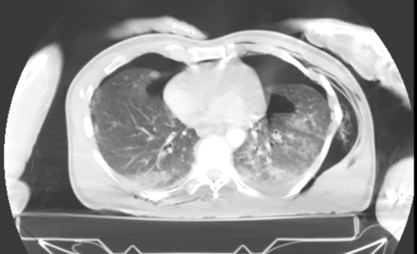
On physical examination, the manubrium sterni appeared anteriorly displaced. Palpation revealed pain, sternal instability, and subcutaneous crepitus. Routine biochemical investigations were unremarkable. Thoracic computed tomography (CT) revealed bilateral multiple rib fractures, bilateral hemopneumothorax, subcutaneous emphysema (Figure 1), and a fracture at the angulus sterni resulting in separation between the manubrium and the corpus sterni (Figure 2).

Figure 1 Axial thoracic CT image demonstrating trauma-induced bilateral hemopneumothorax, multiple rib fractures, and subcutaneous emphysema.
Based on these findings, the patient was taken to surgery after obtaining informed consent and completing preoperative preparation. An anterior approach was used to expose the sternal fracture line intraoperatively. The fracture ends were mobilized, anatomically reduced, and stabilized using a titanium plate (Figure 3). Postoperative chest radiographs confirmed appropriate alignment and stabilization of the sternum (Figure 4). The patient was closely monitored postoperatively; no complications were observed, and he was discharged within the expected recovery period.
Sternal fractures most commonly occur at the anatomical regions of the manubrium or the body of the sternum. Unstable or displaced fractures can influence mortality due to the risk of associated thoracic organ injury. The overall mortality rate associated with sternal fractures is reported to be approximately 0.7%.3 Complications such as pneumothorax, hemothorax, pulmonary contusion, and tracheobronchial injuries require immediate clinical intervention.4
On physical examination, patients with sternal fractures typically present with localized, sharp pain upon palpation. Approximately half of the cases show soft tissue swelling or ecchymosis, and deformity may be detected on manual inspection. Dyspnea is observed in up to 20% of patients. The pain often intensifies with coughing or deep inspiration.
The initial imaging modality in suspected sternal fractures is typically a standard chest radiograph. Lateral chest radiographs offer greater diagnostic value since sternal fractures are usually transverse and any displacement occurs in the sagittal plane. The diagnostic sensitivity of anteroposterior chest radiographs is estimated to be around 50%.5 Computed tomography (CT) remains the most sensitive and accurate imaging tool for the diagnosis of sternal fractures. One study demonstrated that 94% of sternal fractures in thoracic trauma patients were only detectable by chest CT.6
Conservative management is generally appropriate for stable sternal fractures without associated life-threatening complications. Most isolated fractures heal within approximately 10 weeks with analgesia and supportive care.7 However, surgical intervention may be required in displaced or unstable fractures due to their potential impact on cardiopulmonary function. Rarely, associated injuries such as tracheobronchial rupture, esophageal damage, diaphragmatic tears, cardiac trauma, or blunt aortic injury may also necessitate surgical management.8
Sternal fixation can be achieved using stainless steel wire cerclage or more contemporary techniques such as plating systems with screws. Titanium plates are particularly favored due to their strength, biocompatibility, and conformity to anatomical curvature.9
In recent years, treatment of sternal fractures has improved with the use of titanium plating systems, which offer excellent biocompatibility and durability. These implants fit the sternum's curvature well and minimize tissue damage, leading to less postoperative pain and quicker recovery.
Early surgical stabilization improves mobilization, reduces pulmonary complications, and shortens hospital stay. However, surgical indications must be individualized. In polytrauma cases, sternal fractures may indicate additional thoracic injuries, so preoperative cardiac evaluation—including enzymes, ECG, and echocardiography—is essential.
Thoracic CT with multiplanar and 3D imaging helps guide surgery. Early physiotherapy and breathing exercises further reduce complications. Overall, multidisciplinary care is crucial for optimal recovery and outcomes.
Although traumatic sternal fractures are relatively rare, their proximity to vital intrathoracic structures necessitates careful clinical assessment. The severity of the clinical presentation, the presence of associated injuries, and the patient’s overall condition are key factors in determining the appropriate treatment strategy. Isolated, stable fractures generally respond well to conservative management. However, in displaced or unstable fractures, surgical fixation provides superior anatomical alignment and functional outcomes. A surgical approach not only facilitates early mobilization but also significantly improves the patient’s overall quality of life.
None.
Authors declare no conflict of interest.

©2025 Muzoglu, et al. This is an open access article distributed under the terms of the, which permits unrestricted use, distribution, and build upon your work non-commercially.