Clinical Images Volume 5 Issue 2
Traumatic eosinophilic ulcer clinical images
Yousif I Eltohami, Amal H Abuaffan
Regret for the inconvenience: we are taking measures to prevent fraudulent form submissions by extractors and page crawlers. Please type the correct Captcha word to see email ID.

University of Khartoum, Faculty of Dentistry, Sudan
Correspondence: Amal H Abuaffan, University of Khartoum, Faculty of Dentistry, Sudan
Received: September 25, 2015 | Published: October 12, 2016
Citation: Eltohami YI, Abuaffan AH. Traumatic eosinophilic ulcer clinical images. J Dent Health Oral Disord Ther. 2016;5(2):246. DOI: 10.15406/jdhodt.2016.05.00149
Download PDF
Case
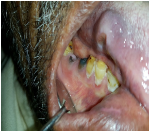
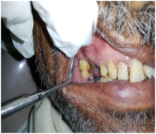
- A Sudanese male 54 years old came to the dental clinic complaining from painful endophytic ulcer within 2 weeks duration.
- On intra oral examination the ulcer had indurated base and everted edges with no discharge, there are sharp badly decayed adjacent right lower premolars. Incisional biopsy showed inflammatory eosinophilic granuloma.

Figure 1 Shows endophytic ulcer beside sharp adjacent posterior teeth.

Figure 2 Shows the relationship between the ulcer & the sharp adjacent posterior teeth in occlusion.
Funding
Acknowledgments
Conflicts of interest
Authors declare that there is no conflict of interest.

©2016 Eltohami, et al. This is an open access article distributed under the terms of the,
which
permits unrestricted use, distribution, and build upon your work non-commercially.


