Journal of
eISSN: 2373-4345


Research Article Volume 1 Issue 5
Department of Preventive Dentistry, University of Benin, Nigeria
Correspondence: Ajayi EO, Orthodontic Unit, Department of Preventive Dentistry, School of Dentistry College of Medical Sciences, University of Benin, Benin City, Nigeria, Tel 802-300-3683
Received: July 27, 2014 | Published: September 22, 2014
Citation: Ajayi EO. Regression equations and probability tables for mixed dentition analysis in a Nigerian population. J Dent Health Oral Disord Ther. 2014;1(4):121-128. DOI: 10.15406/jdhodt.2014.01.00027
Objectives: The objectives of this study were to evaluate the applicability of both the Moyers and the Tanaka and Johnston mixed dentition space analysis in a Nigerian population, and develop a new probability tables and regression equations for prediction of the size of unerupted canines and premolars in Nigerian population.
Methods: The mesiodistal crown dimensions of 54 dental casts of Nigerian subjects were measured with digital calipers. Gender differences were evaluated with independent t-test. Correlation coefficients between the combined mesiodistal widths of the permanent mandibular incisors and the canine and premolars of the maxillary and mandibular arches were determined respectively. Linear regression equations and probability tables were derived and used to compare actual Nigerian tooth widths with those predicted by the Moyer’s probability table and regression equations of Tanaka and Johnston.
Results: The regression equations for the maxillary arch males: Y=0.49x+10.98, females: Y=0.47x+12.95, sexes combined: Y=0.47x+11.49, and the mandibular arch males: Y=0.54x+9.53, females: Y=0.39x+12.75, sexes combined: Y=0.51x+10.27 were derived. No statistically significant differences (p>0.05) were observed in the mesiodistal widths of maxillary canine and premolars for combined sexes at 75th percentile, and in the mandibular arch for male at 85th percentile confidence levels when compared with those of the Moyers’ probability tables, while there were statistically significant differences (p<0.05) at all the remaining percentile levels. Tanaka and Johnston’s equations underestimated canine and premolars mesiodistal widths in the mandibular and maxillary arches.
Conclusion: The Moyers and Tanaka and Johnston mixed dentition space analysis has limited application in this sample of Nigerian population and the newly proposed probability tables would be more accurate for Nigerian subjects.
Keywords: mesiodistal widths, moyer’s probability table, tanaka and johnston regression equations, mixed dentition analysis, nigerian population
The mixed dentition space analysis is an essential part of orthodontic diagnostic procedures required to determine the amount of space available for the alignment of unerupted permanent teeth in a dental arch.1 Space analysis during the mixed dentition stage facilitates the determination of any tooth size to arch length discrepancy and planning of an appropriate orthodontic treatment for the patients. The mixed-dentition arch analysis is therefore an important criterion in determining whether the treatment plan is going to involve serial extraction, guidance of eruption, space maintenance, space regaining, or just periodic observation of the patient.2 Information on the mesiodistal widths of an individual maxillary and mandibular tooth is crucial in the analysis of space requirement and availability in the arches. However, in the mixed dentition stage the determination of tooth size entails accurate prediction of the mesiodistal width of the unerupted permanent canines and premolars in both the maxillary and mandibular arches.
There have been various methods developed for space analysis and prediction of the sizes of unerupted teeth. The three main methods utilized includes direct measurement of teeth from the study models and use of prediction tables and equations,3–5 direct measurement of teeth sizes from radiographs,6–8 and a combination of measurement of tooth sizes on radiographs and use of prediction tables.2,9,10 The combined methods of tooth sizes measurement from the study models and radiographs have been suggested for better accuracy of space prediction even though the radiographic method is more complex and as such it is used infrequently.
The prediction methods of Moyers3,4 and the Tanaka and Johnston5 mixed dentition space analysis have been widely adopted and are most commonly used in clinical practices. The Moyers3 method involved direct measurement of teeth from study models with a generation of data for prediction of the sizes of unerupted canines and premolars from probability table for the sexes combined. Moyers4 also further provided a different probability tables for prediction of the sizes of unerupted canines and premolars for males and females separately. Similarly, Tanaka and Johnston5 developed prediction tables comparable with that of Moyers3 from measurement of teeth from study models and introduced the application of simple regression equations for estimating the summed width of the maxillary and mandibular canine and premolar segments.
The data for the prediction methods of Moyers3,4 and Tanaka and Johnston5 were based on a sample of white children of Northern European descent and the reliability of these prediction methods have been shown among this population.11 However, the validity and accuracy of these data have been questioned when applied to subjects from different ethnic and racial groups since various studies have shown that tooth sizes vary significantly between different population and racial groups.12,13 It is therefore important to determine prediction aids for each racial group since each population has its own tooth characteristics and evaluate possible gender differences and secular trend.14–21 Variation in tooth sizes has also been described in Nigerian subjects when compared to British and African American subjects respectively.22,23 Presently, there is no prior study that has evaluated space analysis in mixed dentition among Nigerian subjects. It is therefore desirable to develop a mixed dentition prediction aids that can be used for space analysis in a Nigerian population.
The purposes of this study therefore were to evaluate the applicability of both the Moyers3,4 and Tanaka and Johnston 5 mixed dentition space analysis among the Nigerian subjects and to construct regression equations and probability tables based on the tooth sizes of Nigerian population sample for more accurate prediction of the size of unerupted canines and premolars in a Nigerian population.
The materials for this study consisted of dental casts of Nigerian subjects who were students at the School of Dentistry of University of Benin, Benin City. The subjects belonged to the predominant ethnic groups of the south-southern and eastern regions of Nigeria. The inclusion criteria were that the subject being a native Nigerian, permanent teeth present and fully erupted, particularly from the first molar to first molar, no missing teeth or abnormally sized or shaped teeth, no interproximal caries or excess tooth materials as a result of restorations and no presence of dental attrition. A sample of 54 subjects (33 males and 21 females) with a mean age of 26.6±2.1 years who met the inclusion criteria was selected. The study was approved by the Research ethics committee of College of Medicine of the University of Benin and the subjects gave their explicit consent to participate.
An alginate impression of upper and lower arches cast in dental stone was made for each subject. The maximum mesiodistal widths of the mandibular permanent central and lateral incisors, mandibular and maxillary permanent canines, first and second premolars were measured on the dental casts with a digital vernier caliper. The caliper was placed parallel to the occlusal plane of each tooth with its sharp points at the greatest distances between the contact points on the proximal surface of each tooth and measurement taken and rounded to the nearest 0.1mm. Each tooth was measured twice and intra-examiner consistency was set at 0.2mm as suggested by Bishara et al.13. New sets of measurements were taken if the differences were greater than this limit and the nearest two measurements obtained were then averaged. The repeat measurements of twenty randomly selected dental casts two weeks after the first measurement indicated good intra-examiner reliability as there was no statistically significant differences between the initial and repeated data.24
Descriptive statistics including means, standard deviations and ranges were calculated for tooth dimensions. A paired t-test was performed to determine if there was any significant statistical difference in the tooth size dimensions between the left and right side of the arch in the maxilla and mandible. An independent t-test was used to determine any statistical gender differences in mesiodistal tooth dimensions.
From the data obtained, correlation coefficients (r) and linear regression equations (y=a+bx) were formulated to evaluate the relationships between the sum of the mesiodistal widths of the four mandibular incisors (x) and the sum of the mandibular and maxillary arch canines and first and second premolars (y). The constants a and b in the standard linear regression equation (y=a+bx), the coefficients of determination (r2), and the standard errors of the estimates (SEE) were calculated for the males, females and both sexes combined. The data derived from this present study were then used to frame prediction equations and probability tables for Nigerian subjects which were then compared with the prediction methods of Moyers 3,4 and the Tanaka and Johnston5 equation. Wilcoxon signed rank test was used to statistically compare the predicted values obtained with the Moyers probability tables 3,4 at the 5th to 95th percentile confidence levels. Statistical significance was regarded when p<0.05. The data entry and analysis was carried out with Statistical Package for Social Sciences software version 16 (SPSS, Chicago, Illinois).
The descriptive statistics for the sums of the mesiodistal crown dimensions of the four mandibular incisors, mandibular canine and premolars and maxillary canine and premolars for the males and females separately and the sexes combined are presented in (Table 1). The comparison of the mean mesiodistal teeth widths of the summed mandibular incisors, mandibular and maxillary canine and premolars among the gender revealed no statistically significant differences even though the males have slightly wider teeth sizes than females (P>0.05). There was also no statistical differences in the tooth dimensions between the left and right sides of the arches (P>0.05).
|
Tooth Group |
Sex |
n (mm) |
Mean (mm) |
Range (mm) |
SD |
|
Mandibular incisor |
M |
33 |
23.54 |
19.66 - 26.10 |
1.50 |
|
Mandibular canine-premolar |
M |
33 |
22.34 |
20.51 - 24.42 |
1.09 |
|
Maxillary canine-premolar |
M |
33 |
22.59 |
20.72 - 25.62 |
1.04 |
|
Mandibular incisor |
F |
21 |
23.43 |
19.76 - 24.90 |
1.20 |
|
Mandibular canine-premolar |
F |
21 |
21.85 |
19.80 - 23.70 |
0.93 |
|
Maxillary canine-premolar |
F |
21 |
22.41 |
19.95 - 24.42 |
1.16 |
|
Mandibular incisor |
M+F |
54 |
23.50 |
19.66 - 26.10 |
1.38 |
|
Mandibular canine-premolar |
M+F |
54 |
22.15 |
19.80 - 24.42 |
1.05 |
|
Maxillary canine-premolar |
M+F |
54 |
22.52 |
19.95 - 25.62 |
1.08 |
Table 1 Descriptive statistics for the sum of mesiodistal widths of mandibular incisors, mandibular and maxillary canines and premolars in a Nigerian sample
M, male; F, female; M + F, combined males and females; SD, standard deviation; n, total number
Table 2 shows the correlation coefficients (r) between the mesiodistal widths of the mandibular incisors and the mandibular and maxillary canines and premolars for males, females and the sexes combined and the regression values a (y-intercept) and b (slope of the regression line) derived from the standard linear regression equation y=a+bx, the SEE (standard error of the estimate) and the coefficient of determination (r2) of the mandibular and maxillary regression equations. The correlation coefficients (r) ranged from 0.42 to 0.75, with a high positive correlations for canine - premolars segment in both mandibular and maxillary for the sexes combined and highest coefficients observed in the male canine-premolars segment in both arches.
|
Regression Constants |
||||||
|
Sex |
Canine-premolar segment |
r |
a |
b |
SEE |
r2 |
|---|---|---|---|---|---|---|
|
Male |
Mandibular |
0.75 |
9.53 |
0.54 |
0.73 |
0.56 |
|
Male |
Maxillary |
0.71 |
10.98 |
0.49 |
0.74 |
0.51 |
|
Female |
Mandibular |
0.50 |
12.75 |
0.39 |
0.83 |
0.25 |
|
Female |
Maxillary |
0.42 |
12.95 |
0.40 |
1.08 |
0.17 |
|
M + F |
Mandibular |
0.67 |
10.27 |
0.51 |
0.79 |
0.44 |
|
M + F |
Maxillary |
0.60 |
11.49 |
0.47 |
0.87 |
0.36 |
Table 2 Regression parameters for prediction of mesiodistal widths of canine and premolars in the mandibular and maxillary buccal segments in a Nigerian sample
M + F, combined males and females; r, correlation coefficient; regression constants, a and b; SEE: standard error of estimate; r2, coefficients of determination
The data derived from the regression analysis were then used to formulate new prediction equations that can be used clinically to predict the mesiodistal crown widths of the unerupted canine and premolars (y) in both arches among Nigerian population when the mesiodistal crown widths of the four mandibular incisors (x) are known among the males, females and with the sexes combined as shown in (Table 3).
|
Population Group |
Sex |
Arch |
Regression Equation Y = a+bx |
|
Nigerian |
Male |
Mandibular |
Y = 9.53 + 0.54x |
|
Male |
Maxillary |
Y = 10.98 + 0.49x |
|
|
Female |
Mandibular |
Y = 12.75 + 0.39x |
|
|
Female |
Maxillary |
Y = 12.95 + 0.40x |
|
|
M + F |
Mandibular |
Y = 10.27 + 0.51x |
|
|
M + F |
Maxillary |
Y = 11.49 + 0.47x |
Table 3 Regression equations for prediction of summation of mesiodistal widths of canine and premolars in the mandibular and maxillary buccal segments in a Nigerian sample
M + F, combined males and females
Table 4 and 5 show the proposed new probability tables for Nigerian population prepared from the regression equation of this present study patterned after the Moyer’s probability table for combined sexes to predict the mesiodistal crown widths of the unerupted canine and premolars in both the mandible and the maxilla respectively, while the probability tables for males and females separately are shown in (Table 6 and 7).
|
Percentile |
20.5 |
21 |
21.5 |
22 |
22.5 |
23 |
23.5 |
24 |
24.5 |
25 |
25.5 |
26 |
26.5 |
27 |
|
5% |
20.66 |
20.91 |
21.16 |
21.42 |
21.67 |
21.92 |
22.18 |
22.43 |
22.68 |
22.94 |
23.19 |
23.44 |
23.69 |
23.95 |
|
15% |
20.71 |
20.96 |
21.21 |
21.47 |
21.72 |
21.97 |
22.23 |
22.48 |
22.73 |
22.99 |
23.24 |
23.49 |
23.74 |
24.00 |
|
25% |
20.76 |
21.01 |
21.26 |
21.52 |
21.77 |
22.02 |
22.28 |
22.53 |
22.78 |
23.04 |
23.29 |
23.54 |
23.79 |
24.05 |
|
35% |
20.81 |
21.06 |
21.31 |
21.57 |
21.82 |
22.07 |
22.33 |
22.58 |
22.83 |
23.08 |
23.34 |
23.59 |
23.84 |
24.10 |
|
50% |
20.88 |
21.13 |
21.39 |
21.64 |
21.89 |
22.15 |
22.40 |
22.65 |
22.91 |
23.16 |
23.41 |
23.66 |
23.92 |
24.17 |
|
65% |
20.96 |
21.21 |
21.46 |
21.71 |
21.97 |
22.22 |
22.47 |
22.73 |
22.98 |
23.23 |
23.48 |
23.74 |
23.99 |
24.24 |
|
75% |
21.01 |
21.26 |
21.51 |
21.76 |
22.02 |
22.27 |
22.52 |
22.78 |
23.03 |
23.28 |
23.53 |
23.79 |
24.04 |
24.29 |
|
85% |
21.05 |
21.31 |
21.56 |
21.81 |
22.07 |
22.32 |
22.57 |
22.83 |
23.08 |
23.33 |
23.58 |
23.84 |
24.09 |
24.34 |
|
95% |
21.10 |
21.36 |
21.61 |
21.86 |
22.12 |
22.37 |
22.62 |
22.88 |
23.13 |
23.38 |
23.63 |
23.89 |
24.14 |
24.39 |
Table 4 Probability table for predicting the mesiodistal widths of unerupted canine and premolars in the mandibular buccal segment in a Nigerian population for combined males and females
|
Percentile |
20.5 |
21 |
21.5 |
22 |
22.5 |
23 |
23.5 |
24 |
24.5 |
25 |
25.5 |
26 |
26.5 |
27 |
|
5% |
21.14 |
21.37 |
21.61 |
21.84 |
22.08 |
22.31 |
22.55 |
22.78 |
23.02 |
23.25 |
23.49 |
23.72 |
23.96 |
24.19 |
|
15% |
21.19 |
21.43 |
21.66 |
21.90 |
22.13 |
22.37 |
22.60 |
22.84 |
23.07 |
23.31 |
23.54 |
23.78 |
24.01 |
24.24 |
|
25% |
21.25 |
21.48 |
21.72 |
21.95 |
22.18 |
22.42 |
22.65 |
22.89 |
23.12 |
23.36 |
23.59 |
23.83 |
24.06 |
24.30 |
|
35% |
21.30 |
21.53 |
21.77 |
22.00 |
22.24 |
22.47 |
22.71 |
22.94 |
23.18 |
23.41 |
23.65 |
23.88 |
24.12 |
24.35 |
|
50% |
21.38 |
21.61 |
21.85 |
22.08 |
22.32 |
22.55 |
22.79 |
23.02 |
23.26 |
23.49 |
23.73 |
23.96 |
24.20 |
24.43 |
|
65% |
21.46 |
21.69 |
21.93 |
22.16 |
22.40 |
22.63 |
22.87 |
23.10 |
23.34 |
23.57 |
23.81 |
24.04 |
24.27 |
24.51 |
|
75% |
21.51 |
21.75 |
21.98 |
22.22 |
22.45 |
22.68 |
22.92 |
23.15 |
23.39 |
23.62 |
23.86 |
24.09 |
24.33 |
24.56 |
|
85% |
21.56 |
21.80 |
22.03 |
22.27 |
22.50 |
22.74 |
22.97 |
23.21 |
23.44 |
23.68 |
23.91 |
24.15 |
24.38 |
24.62 |
|
95% |
21.62 |
21.85 |
22.09 |
22.32 |
22.56 |
22.79 |
23.03 |
23.26 |
23.49 |
23.73 |
23.96 |
24.20 |
24.43 |
24.67 |
Table 5 Probability table for predicting the mesiodistal widths of unerupted canine and premolars in the maxillary buccal segment in a Nigerian population for combined males and females
Percentile |
19.5 |
20 |
20.5 |
21 |
21.5 |
22 |
22.5 |
23 |
23.5 |
24 |
24.5 |
25 |
25.5 |
|
Males |
5% |
20.16 |
20.44 |
20.71 |
20.98 |
21.25 |
21.52 |
21.80 |
22.07 |
22.34 |
22.61 |
22.88 |
23.16 |
23.43 |
15% |
20.21 |
20.48 |
20.75 |
21.03 |
21.30 |
21.57 |
21.84 |
22.11 |
22.39 |
22.66 |
22.93 |
23.20 |
23.47 |
|
25% |
20.27 |
20.54 |
20.81 |
21.08 |
21.35 |
21.63 |
21.90 |
22.17 |
22.44 |
22.71 |
22.99 |
23.26 |
23.53 |
|
35% |
20.30 |
20.57 |
20.84 |
21.12 |
21.39 |
21.66 |
21.93 |
22.20 |
22.48 |
22.75 |
23.02 |
23.29 |
23.56 |
|
50% |
20.37 |
20.64 |
20.91 |
21.19 |
21.46 |
21.73 |
22.00 |
22.27 |
22.55 |
22.82 |
23.09 |
23.36 |
23.63 |
|
65% |
20.44 |
20.71 |
20.98 |
21.25 |
21.53 |
21.80 |
22.07 |
22.34 |
22.61 |
22.89 |
23.16 |
23.43 |
23.70 |
|
75% |
20.48 |
20.76 |
21.03 |
21.30 |
21.57 |
21.84 |
22.12 |
22.39 |
22.66 |
22.93 |
23.20 |
23.48 |
23.75 |
|
85% |
20.53 |
20.80 |
21.07 |
21.34 |
21.62 |
21.89 |
22.16 |
22.43 |
22.70 |
22.98 |
23.25 |
23.52 |
23.79 |
|
95% |
20.57 |
20.85 |
21.12 |
21.39 |
21.66 |
21.93 |
22.21 |
22.48 |
22.75 |
23.02 |
23.29 |
23.57 |
23.84 |
|
|
||||||||||||||
Females |
5% |
20.76 |
20.86 |
20.97 |
21.07 |
21.18 |
21.28 |
21.39 |
21.50 |
21.60 |
21.71 |
21.81 |
21.92 |
22.02 |
15% |
20.82 |
20.92 |
21.03 |
21.13 |
21.24 |
21.34 |
21.45 |
21.56 |
21.66 |
21.77 |
21.87 |
21.98 |
22.08 |
|
25% |
20.88 |
20.98 |
21.09 |
21.19 |
21.30 |
21.41 |
21.51 |
21.62 |
21.72 |
21.83 |
21.93 |
22.04 |
22.15 |
|
35% |
20.94 |
21.04 |
21.15 |
21.26 |
21.36 |
21.47 |
21.57 |
21.68 |
21.78 |
21.89 |
22.00 |
22.10 |
22.21 |
|
50% |
21.03 |
21.14 |
21.24 |
21.35 |
21.45 |
21.56 |
21.66 |
21.77 |
21.88 |
21.98 |
22.09 |
22.19 |
22.30 |
|
65% |
21.12 |
21.23 |
21.33 |
21.44 |
21.55 |
21.65 |
21.76 |
21.86 |
21.97 |
22.07 |
22.18 |
22.29 |
22.39 |
|
75% |
21.18 |
21.29 |
21.39 |
21.50 |
21.61 |
21.71 |
21.82 |
21.92 |
22.03 |
22.13 |
22.24 |
22.35 |
22.45 |
|
85% |
21.24 |
21.35 |
21.46 |
21.56 |
21.67 |
21.77 |
21.88 |
21.98 |
22.09 |
22.20 |
22.30 |
22.41 |
22.51 |
|
95% |
21.31 |
21.41 |
21.52 |
21.62 |
21.73 |
21.83 |
21.94 |
22.05 |
22.15 |
22.26 |
22.36 |
22.47 |
22.57 |
|
Table 6 Probability table for predicting the mesiodistal widths of unerupted canine and premolars in the mandibular buccal segment in a Nigerian population for males and females separately
Percentile |
19.5 |
20 |
20.5 |
21 |
21.5 |
22 |
22.5 |
23 |
23.5 |
24 |
24.5 |
25 |
25.5 |
|
Males |
5% |
20.62 |
20.87 |
21.12 |
21.36 |
21.61 |
21.86 |
22.10 |
22.35 |
22.60 |
22.84 |
23.09 |
23.34 |
23.58 |
15% |
20.67 |
20.92 |
21.17 |
21.41 |
21.66 |
21.91 |
22.15 |
22.40 |
22.65 |
22.89 |
23.14 |
23.39 |
23.63 |
|
25% |
20.72 |
20.97 |
21.22 |
21.46 |
21.71 |
21.96 |
22.20 |
22.45 |
22.70 |
22.94 |
23.19 |
23.44 |
23.68 |
|
35% |
20.78 |
21.02 |
21.27 |
21.52 |
21.76 |
22.01 |
22.26 |
22.50 |
22.75 |
23.00 |
23.24 |
23.49 |
23.74 |
|
50% |
20.85 |
21.10 |
21.34 |
21.59 |
21.84 |
22.08 |
22.33 |
22.58 |
22.82 |
23.07 |
23.32 |
23.56 |
23.81 |
|
65% |
20.93 |
21.17 |
21.42 |
21.67 |
21.91 |
22.16 |
22.41 |
22.65 |
22.90 |
23.15 |
23.39 |
23.64 |
23.89 |
|
75% |
20.98 |
21.23 |
21.47 |
21.72 |
21.96 |
22.21 |
22.46 |
22.70 |
22.95 |
23.20 |
23.44 |
23.69 |
23.94 |
|
85% |
21.03 |
21.28 |
21.52 |
21.77 |
22.02 |
22.26 |
22.51 |
22.76 |
23.00 |
23.25 |
23.50 |
23.74 |
23.99 |
|
95% |
21.08 |
21.33 |
21.57 |
21.82 |
22.07 |
22.31 |
22.56 |
22.81 |
23.05 |
23.30 |
23.55 |
23.79 |
24.04 |
|
|
||||||||||||||
Females |
5% |
20.85 |
21.05 |
21.26 |
21.46 |
21.66 |
21.86 |
22.06 |
22.26 |
22.47 |
22.67 |
22.87 |
23.07 |
23.27 |
15% |
20.91 |
21.11 |
21.32 |
21.52 |
21.72 |
21.92 |
22.12 |
22.32 |
22.53 |
22.73 |
22.93 |
23.13 |
23.33 |
|
25% |
20.97 |
21.17 |
21.37 |
21.58 |
21.78 |
21.98 |
22.18 |
22.38 |
22.59 |
22.79 |
22.99 |
23.19 |
23.39 |
|
35% |
21.03 |
21.23 |
21.43 |
21.64 |
21.84 |
22.04 |
22.24 |
22.44 |
22.65 |
22.85 |
23.05 |
23.25 |
23.45 |
|
50% |
21.12 |
21.32 |
21.52 |
21.73 |
21.93 |
22.13 |
22.33 |
22.53 |
22.74 |
22.94 |
23.14 |
23.34 |
23.54 |
|
65% |
21.21 |
21.41 |
21.61 |
21.82 |
22.02 |
22.22 |
22.42 |
22.62 |
22.82 |
23.03 |
23.23 |
23.43 |
23.63 |
|
75% |
21.27 |
21.47 |
21.67 |
21.87 |
22.08 |
22.28 |
22.48 |
22.68 |
22.88 |
23.09 |
23.29 |
23.49 |
23.69 |
|
85% |
21.33 |
21.53 |
21.73 |
21.93 |
22.14 |
22.34 |
22.54 |
22.74 |
22.94 |
23.15 |
23.35 |
23.55 |
23.75 |
|
95% |
21.39 |
21.59 |
21.79 |
21.99 |
22.20 |
22.40 |
22.60 |
22.80 |
23.00 |
23.21 |
23.41 |
23.61 |
23.81 |
|
Table 7 Probability table for predicting the mesiodistal widths of unerupted canine and premolars in the maxillary buccal segment in a Nigerian population for males and females separately
There are no statistically significant differences (P>0.05) between the predicted values for unerupted canine and premolars in the maxilla for combined sexes at 75% and in the mandibular arch for males at 85% confidence levels compared with those of Moyers probability table where prediction of the actual mesiodistal width of canines and premolar in Nigerian subjects are possible. However, there is highly significant differences (P<0.01) at all other percentile confidence levels except in the mandible for females at 95% and also in the maxilla for males and females at 95% respectively (P<0.05).
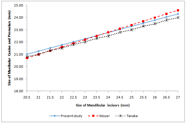
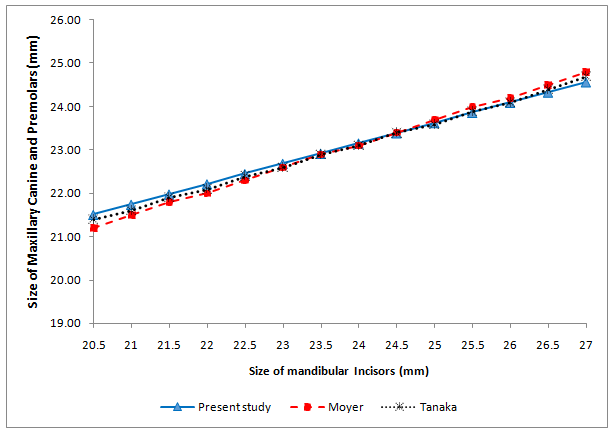
Graphical comparison between the predicted values from the proposed equations of the Nigerian subjects and that of the Tanaka and Johnston equations and the Moyers probability charts at the 75th percentile level for the maxillary and mandibular canine-premolar segments for both sexes combined are depicted in (Figure 1 & 2). Figure 1 shows that the Tanaka and Johnston values entirely underestimated mesiodistal width sizes of mandibular canine and premolars in Nigerian subjects. The Moyers chart also underestimated mandibular canine and premolars sizes when the sum of the total mandibular incisors is less than 23mm and also predicted larger sizes for Nigerian subjects when the sum of the total mandibular incisors is above 25mm. However, the predicted Nigerian sizes for canine and premolars and that of Moyers are approximately the same when the sum of the total mandibular incisors is between 23mm and 25mm. Figure 2 illustrates that the Moyers and the Tanaka and Johnston values were closely related to the Nigerian predicted values for maxillary canine and premolars when the total mandibular incisors is between 23mm and 25mm but underestimated mesiodistal width sizes of canine and premolars in Nigerian subjects when the total mandibular incisors is less than 23mm.


The application of regression equations and probability charts in estimating the mesiodistal widths of the unerupted permanent canines and premolars during mixed dentition was deployed among this sample of Nigerian subjects. This study revealed no statistically significant gender differences in the mesiodistal crown dimensions of the mandibular incisors, mandibular and maxillary canines and premolars even though the males have a slightly wider teeth sizes than females. The mean mesiodistal crown dimensions for males, females and both sexes combined were reported to facilitate comparison with studies from other racial and ethnic groups which gave only sex-specific data since sexual dimorphism in teeth sizes has been reported in some studies. 16,17,19,25 The combined gender mean values obtained for the mandibular incisors, mandibular and maxillary canines and premolars were similar to reported findings among the Black Americans, Thai, Senegalese and Asian-Americans populations.15,17,25,26 Sex-specific values revealed higher values among Nigerian subjects compared to Nepalese populations21 and the teeth sizes are also comparable to the odontometric data reported for Black South Africans14 who however have slightly larger teeth sizes among the males. This observed variation in tooth sizes in different racial groups further buttresses the need for development of prediction aids for each racial group. The causes of tooth-size variations in different racial groups have been attributed possibly to genetic, nutrition and environmental factors.
The correlation coefficients between the summed widths of mandibular incisors and the canine and premolars obtained in this study for both the mandibular and maxillary arches in the males were high 0.71 and 0.75 respectively indicating a high predictability in determining the mesiodistal width of unerupted canine and premolars. The correlation coefficients for combined sexes was also high and further reinforce the relativeness of mandibular incisors as a predictor aid to determine the widths of unerupted canine and premolars for this sample of Nigerian population. The correlation coefficients obtained in this study is comparable to high correlation coefficients in ranges of 0.6 and above reported among American White children5 Black Americans15 Thai17 and Senegalese populations 25 with predominance of higher values in males as also reported in other studies.16,19,25 However, lower values for correlation coefficients were reported in males among the Thai17 and Nepalese21 populations possibly due to gender variability in these populations. The coefficient of determination r2 of the range of 36% to 44% was observed among the sexes revealing the total variance in canine-premolar widths that could be determined form the knowledge of the combined widths of mandibular incisors. Similar values were obtained in Thai and Pakistan populations.17,27 Discrepancy in the values of coefficient of determination was also observed among the genders with higher values among the males as also reported in other populations19,25 and lower values in females which could possibly be attributed to sample size. The standard error of estimate SEE was 0.79 to 0.87 for combined sexes, these lower values indicate better prediction equation and similar to values reported in other populations.17,27
This study shows that the Tanaka and Johnston values underestimated mesiodistal width sizes of mandibular canine and premolars in Nigerian subjects and also predicted lower values than actual width of the canine and premolars in the maxillary arch when the sum of the total mandibular incisors is less than 23mm. However, the Tanaka and Johnston equations and the Moyers probability chart gave predictions that were closely related to the Nigerian actual values for canine and premolars when the total mandibular incisors is greater than 23mm in the maxillary arch with a tendency towards slight overestimation as summed total mandibular incisors sizes increase. Similarly, Moyers probability chart also predicted lower values for the actual mandibular and maxillary canine and premolars widths when the sum of the total mandibular incisors is less than 23mm in both arches as observed with the Tanaka and Johnston equation at the 75th percentile levels. There were no statistically significant difference between actual mesiodistal width of canines and premolars in Nigerian sample and the predicted width from the Moyers charts only at the recommended 75th percentile in the maxillary arch for both sexes combined and also in the mandibular arch for male at 85% percentile confidence levels (P>0.05) while there were significant differences at the remaining percentiles. Similar inaccurate predictions with a possibility of overestimation or underestimation of the actual width of the canine and premolars with the Tanaka and Johnston’s equations and Moyer’s probability chart have also been reported in Black South African,14 Indian,16 Saudi Arab,18 Iranian,20 Nepalese,21 Senegalese,25 Jordanian28 and Turkish populations.29 It thus reflects the limitations of these methods when applied to a sample of population other than European descent because of variability in tooth widths.
The importance of accurate prediction of unerupted tooth sizes can therefore not be overemphasized as the determination of the amount of space available for the accommodation of unerupted permanent teeth in a dental arch will enhance extraction decision or space gaining procedure to be implemented during orthodontic treatment planning for the patients. The proposed new probability tables for males, females and sexes combined for Nigerian population developed from the regression equations of the actual tooth widths of Nigerian sample is therefore recommended in application of mixed dentition analysis in Nigerian population.
This study revealed limited application of the Moyers and the Tanaka and Johnston mixed dentition space analysis in a Nigerian population. A regression equation and new probability tables for prediction of unerupted canine and premolars in the mandibular and maxillary arches for males, females and sexes combined for Nigerian population were developed. Further validation of these mixed dentition prediction aids in a larger sample of Nigerian population is imperative and recommended.
None.
None.
The authors declare that there is no conflict of interest.

©2014 Ajayi. This is an open access article distributed under the terms of the, which permits unrestricted use, distribution, and build upon your work non-commercially.