Journal of
eISSN: 2373-4345


Research Article Volume 3 Issue 3
1Department of Public Health Sciences, Texas A&M University Baylor College of Dentistry, USA
2Undergraduate student, Texas A&M University Baylor College of Dentistry, USA
3Department of Restorative Sciences, Texas A&M University Baylor College of Dentistry, USA
Correspondence: Amal Noureldin, Department of Public Health Sciences, Texas A&M University Baylor College of Dentistry, USA, Tel 214-828-8354, Fax 214-874-4555
Received: November 24, 2015 | Published: December 7, 2015
Citation: Noureldin A, Jupta H, Hunyh T, et al. Re-hardening effect of two-applications of combined fluoride-laser on incipient-enamel lesions. J Dent Health Oral Disord Ther. 2015;3(3):295-298. DOI: 10.15406/jdhodt.2015.03.00088
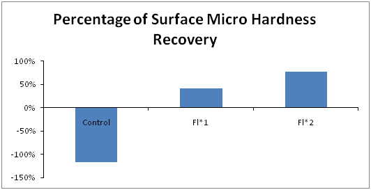
Previous research demonstrated that combined treatment of fluoride followed by laser irradiation propitiates an expressive fluoride uptake, reducing the progression of white-spot-lesions. Since no other studies investigated the treatment effect of repeated applications of fluoride-laser combined treatment, this study aimed to investigate if two-applications of fluoride-laser sequence would re-harden enamel surface of incipient-enamel lesions more than one-time application. 36 enamel slabs (3mm x 3mm x 4mm) were cut from 6 human molars, ground flat, polished and coated with nail varnish except 2x3 windows. White-spot lesions (WSL) were created in all specimens (demineralizing solution/16 hrs). Specimens randomly assigned into 3 groups; (1) (Control) received no treatment, (2) (FL1) one-application of MI fluoride-varnish followed by CO2 laser (short-pulsed 10.6μm, 2.4J/cm2, 10HZ, 10sec), (3) (FL2) two-applications of MI varnish-CO2 laser, specimens were left in distilled water for one day between applications. 8-day pH cycle (2hr demin/ 22hr remin) was carried out for all tested groups. Knoop surface-microhardness using 50-grams/10 seconds (SMH) was measured at baseline, after WSL formation, and after treatment. Percentage of surface microhardness recovery (%SMHR) was calculated. ANOVA followed by Duncan’s Multiple Range Test were used for data analysis (5% significance level). Findings suggest that treating WSL with fluoride-laser sequence was capable of inhibiting further progression and re-hardened incipient-enamel lesions when compared to control (-117%), which showed significant, further decrease in SMH when challenged by pH-cycle. Although two-time application of fluoride-laser showed the highest percentage of SMH recovery (77%), results revealed that it does not provide a significant additional remineralization potential when compared to one-time application (40%).
Keywords: fluoride, lasers, enamel caries, remineralization
CO2 lasers had shown great potential in increasing the resistance of enamel surface to acid attacks. The thermal effect of certain laser parameters proved to cause some structural and chemical changes in enamel. The available data revealed a promising combined treatment when fluoride and laser were used together compared to laser or fluoride treatment alone.
Liu et al.1 explained the possible cariostatic mechanisms of the combined fluoride-laser treatment. They suggested that a laser-induced purification of the human enamel hydroxy apatite structure takes place and plays the major role in this cariostatic process. They also mentioned that the low-energy laser treatment has a photo thermal effect that may cause a reduction in enamel permeability. They also reported an increase in the fluoride uptake by the enamel surface in the form of calcium fluoride. And subsequently, in the presence of fluoride and laser treatment a transformation of hydroxy apatite into “fluoro apatite” takes place, which is more resistant to acid attacks.
Several researchers have evaluated caries prevention using combined lasers and fluoride treatment. However, only minimal research has focused on testing the re-hardening effect on already existing white spot lesions. They always tested one laser effect with one acid attack. But based on previous studies we had the insight of investigating the frequency of laser irradiation combined with fluoride treatment. This study was designed to test the null hypothesis that repeated application of the combined varnish and laser treatment would not have any significant effect on the further progression of the white spot lesion in enamel.
Experimental design
36 enamel slabs were cut from 6 human molars. Following white-spot lesions (WSL) creation in all specimens, they were randomly assigned into 3 groups; (1) (Control) received no treatment, (2) (FL1) one-application of MI fluoride-varnish followed by CO2 laser (short-pulsed 10.6μm, 2.4J/cm2, 10HZ, 10sec), (3) (FL2) two-applications of MI varnish-CO2 laser. Treatments were followed by caries challenge (pH-cycling). The response variable was surface microhardness (SMH), which was measured at baseline, after WSL formation, and after treatment.
Sample preparation
0.1% (wt/vol) thymol solution at 4°C was used for human teeth storage, until the beginning of the experiment.2 Teeth were checked for restorations, cracks, caries or developmental defects. Teeth with intact buccal enamel surfaces were used. Roots were removed using a high-speed hand piece with copious amount of water. Each tooth crown was sectioned to create 3 specimens, using one in each group. To minimize variations in results, the control and the experimental specimens were from the same tooth. The enamel surfaces were fixed in Teflon matrices using casting wax,3 and were ground flat and polished with carbide paper (600, 800 and 1200 grid in sequence) under copious running water on a grinding and polishing machine (DP-9U2; Struers S/A, Copenhagen, Denmark). An acid-resistant nail varnish (Revlon Cherry color) double coated the specimens except for a treatment window (2.0 x 3.0mm) that left exposed. Specimens were stored in distilled water.
Artificial caries lesions
Following a demineralization protocol from Queiroz et al.,4 early caries lesions were created in all groups by individually immersing the specimens in falcon tube containing 12ml demineralizing solution (2ml/mm2 of the enamel area) without agitation at 37% for 64 hours. The demineralization solution composed of 50mM acetate buffer solution containing (1.28mM) calcium nitrate trihydrate, (0.74mM) sodium dihydrogen phosphate monohydrate, and 0.03μg F/mL (0.03 ppm fluoride).The addition of low fluoride concentration (0.03μg F/mL) was to help preserve the enamel surface. This is a relevant aspect when considering the formation of a typical subsurface lesion Rehder Neto et al.5 Then, all specimens were cleaned with a piece of gauze soaked in deionized water and kept in artificial saliva in an incubator at 37°C for 24 hours to be treated later. Artificial saliva formulation consisted of hydrogen carbonate (22.1mmol/L), potassium (16.1mmol/L), sodium (14.5mmol/L), calcium (0.2mmol/L), hydrogen phosphate (2.6mmol/L), boric acid (0.8mmol/L), calcium (0.7mmol/L), thiocyanate (0.2mmol/L) and magnesium (0.2mmol/L) with a pH between 7.4 and 7.8.4
Surface treatment
Following WSL creation, the randomly assigned specimens received the following treatments; (CON) control group: no surface treatment and enamel received pH cycle. (F-L1): One-time application of Fluoride varnish-laser group. Specimens were dried with absorbent paper then MI varnish (5% sodium fluoride varnish with Recaldent (CPP-ACP), GC America, USA) was applied to the treatment window using the application micro brush as directed by the manufacturer. Fluoride-treated surface was irradiated with 10.6μm CO2 laser (Azuryt CTL 1401, CO2 North American Clinical Lazer System, LTD) (fluence per pulse from 3.3 to 4.4 J/cm2, wave length 10.6μm, pulse duration 20μs, pulse repetition rate 20Hz, beam diameter of focus 1100μm). A straight hand piece was used to deliver the laser beam from a distance of approximately 5 mm. Only one operator treated enamel windows with laser in a scanning mode moving the hand piece uniformly and longitudinally over the treatment window. After 4 min, specimens were immersed in artificial saliva at 37°C. After 24hours storage, a knife blade was used to remove the fluoride varnish from enamel surface to resemble the varnish removal in vivo by tooth brushing.3 Then specimens were rinsed with deionized water before the pH-cycle. (F-L2): Two applications of Fluoride varnish-laser: This group received similar treatment as F-L1 group. A second application of the fluoride-laser treatment was carried out at the end of the first pH cycle followed by another 9-day pH cycling.
Artificial cariogenic challenge
After treatment, groups were subjected to a 9-days pH-cycling model (8+1 day remineralization bath at 37°C), following Queiroz protocol.4 All specimens were covered with pink wax except for the treatment window, attached to a piece of orthodontic wire to suspend it in plastic falcon tubes which were kept in an incubator at 37°C and under constant agitation at 200rpm during the whole pH-cycle. The specimens in all groups were immersed for 4h in 25mL demineralization solution (1.28mM calcium nitrate, 0.74mM sodium dihydrogen phosphate, 0.05 M acetate buffer, 0.03μg F/ml, pH 5.0).Followed by thorough rinsing of the specimens (10s) in distilled water and drying with absorbent paper. Then, specimens immersed 20h in 12.5mL remineralization bath (1.5mM calcium nitrate, 0.9mM sodium dihydrogen phosphate, 150mM potassium chloride, 0.1 M Tris buffer, 0.05μg F/ml, pH 7). After 8 days of cycling, remineralization for 24h took place in the 9th day. On the 4th day, the de- and remineralizing solutions were replaced by fresh solution. The plastic falcon tubes with the suspended specimens were kept in an incubator at 37°C and under constant agitation at 200rpm during the whole pH-cycle. After completion of the pH-cycling specimens were stored on wet cotton fabric at room temperature and 100% relative humidity.6
Surface microhardness analysis
1200 grid carbide paper was used to obtain polished, smooth and unscratched enamel. All specimens were tested for (SMH) using 50-gram load for 10seconds. SMH was recorded three times for each specimen, baseline SMH, SMH after induction of WSL, SMH after pH cycling. Five clear flawless indentations spaced 100µm were made at the center of the working enamel surface. The average of the five readings was calculated for each specimen as the microhardness value. Then the percentage of mineral recovery of the SMH values (%SMHR) was calculated by this formula
ANOVA followed by Duncan’s Multiple Range Test were used for data analysis (5% significance level).
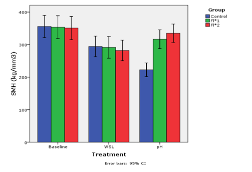
Table 1& Figure 1 shows the descriptive statistics for the three different groups with the different SMH readings Tests detect significant differences at the level of (P≤.001), between various surface treatments at different phases of study. Figure 2 shows the percentage of surface micro hardness recovery for Con, FL1, FL2 groups (-117%, 77%, 40% respectively). Control group was significantly lower compared to FL1 and FL2.
|
Control |
Fl*1 |
Fl*2 |
Baseline |
355.02 (±53.93)a |
353.50 (±55.52)a |
351.10 (±55.81)a |
WSL |
294.26(±50.09)b,c |
291.57 (±52.52)b,c |
281.95 (±50.01)c |
pH |
222.53(±33.25)d |
316.49 (±45.91)a,b,c |
334.94 (±44.68)a,b |
Table 1 Surface Microhardness (SMH) comparison of different surface treatment groups (Mean±SD) at different phases of the study
a, b, c, d: Means with same superscript do not differ each other (Duncan’s Multiple Range Test)

Figure 1 Bar chart of Surface Microhardness (SMH) comparison of different surface treatment groups (Mean±SD) at different phases of the study.

Figure 2 N=57; Epidemiological distribution of the pathological fractures, traumatic fractures, and nonunion.
In our study, we have chosen the CPP-ACP, which is a relatively new mineralization technology. The formula is based on casein phosphopeptide (milk protein casein).5 The CPP (Casein phosphopeptide) is able to stabilize calcium phosphate in nano complexes like ACP (amorphous calcium phosphate). CPP binds to ACP in meta stable solution, which prevents of dissolution of calcium and phosphate ions. By this mechanism CPP-ACP acts as reservoir of bio-available calcium and phosphate. The solutions around the teeth will remain supersaturated thus facilitating remineralization.
The selection of CO2 laser in our study was based on findings of other studies reporting that the CO2 lasers are the most efficient in caries inhibition compared to other lasers.5-9 This could be attributed to the scientific fact that because of the phosphate, carbonate, and hydroxyl groups in the crystalline structure of enamel, dentin, and cementum they have absorption bands in the infrared region (9.0 to 11.0μm region).10,11 These absorption bands are close to the CO2 laser irradiation.10,12-17 This is why these tissues can efficiently absorb the irradiation from the CO2 laser.
In our study the sequence of fluoride followed by laser was selected over laser followed by fluoride. This selection was based on the findings of several studies that reported better acid-resistance of enamel when the first sequence was used.5,7,15,18,19
The one-time and two-time applications of fluoride varnish followed by laser showed statically significant increased SMH values compared to control group (CON) and fluoride-treated group (FV) after the pH challenge. This would translate into increased surface hardness and acid-resistance of the F-L-treated enamel surface. These results were consistent with other in vitro studies that have shown that combined laser-fluoride have beneficial effects on enamel microhardness.5-8,16,20 This re-hardening effect could be attributed to the physico-chemical changes that have been shown to take place after F-L treatment in several studies as increased micro-porosities in tooth structure, increase in deposition of calcium fluoride on surface, partial conversion of hydroxy apatite to fluoro apatite which becomes trapped in the surface and subsurface enamel and crystal growth related to the temperature change.1
The two applications of F-L treatment did not show significant difference compared to one-time application. However, SMH numerical values of the F-L2 group were greater than those in the F-L1 group. This might suggest a beneficial value of repeated application and may be increased hardness of the soft WSL. This might be attributed to the possibility that post lasing the surface twice there was a greater affinity for calcium, phosphate, and fluoride ion and an enhanced accumulation of these minerals.21
In this study, the internal comparison between the experimental treatment and the respective control carried out here in helped eliminate experimental variability with regard to the employed human enamel substrate. One limitation is that it is not possible to predict the further effect of acid attacks since we cannot reflect on the long-term durability of this therapy.
In this vitro study the synergistic effect of fluoride and CO2 laser was confirmed. It showed the ability of the fluoride-laser sequence to treat, re-harden the WSL, and increases the resistance to further acid dissolution. Further studies that simulate the clinical conditions are needed to test optimal frequency and longevity of applications of this combined treatment for the WSL.
The authors would like to express their gratitude to Dr. Jeffrey Rossmann for allowing the use of the CO2 laser.
The author declares that there was no conflict of interest.

©2015 Noureldin, et al. This is an open access article distributed under the terms of the, which permits unrestricted use, distribution, and build upon your work non-commercially.