Journal of
eISSN: 2373-4345


Research Article Volume 3 Issue 2
1Associate Professor of Endodontics, Department of Endodontics, Faculty of Oral and Dental Medicine, Cairo University, Egypt and Senior Consultant of Endodontics, Ministry of Health, Kingdom of Bahrain
2Lecturer of Endodontics, Department of Endodontics, Faculty of Oral and Dental Medicine, Cairo University, Egypt
Correspondence: Mohammad Hamdi Atteia, Department of Endodontics, Associate Professor of Endodontics, Faculty of Oral and Dental Medicine, Cairo University, Egypt and Senior Consultant of Endodontics, Ministry of Health, Kingdom of Bahrain, Tel 2-01115349093, Fax 973-34028896
Received: October 01, 2015 | Published: November 27, 2015
Citation: Atteia MH, Saba AA. Post-treatment radiographic and clinical evaluation of matched-taper single-cone versus warm vertical compaction technique: a one-year follow up study. J Dent Health Oral Disord Ther. 2015;3(2):290-293. DOI: 10.15406/jdhodt.2015.03.00087
Objectives: This study evaluated the long-term post-treatment response of two root canal filling techniques.
Materials and methods: Thirty-two mandibular first molars in patients diagnosed with pulp-periapical pathosis were instrumented and filled with either: (1) Matched-taper single- cone technique using Pro Taper gutta-percha or (2) Warm vertical compaction technique with gutta-percha. AH Plus sealer was used in both groups. Periradicular alveolar bone density of the preoperative radiographs was compared to one-year postoperative recall radiographs using digital x-ray software. One-year postoperative subjective and objective pain assessments were evaluated and pain index was formulated.
Results: Matched-taper single-cone technique showed a higher change in bone density (-1.67) than the warm vertical compaction technique (+0.99), a difference that was statistically non- significant. Gutta-percha warm vertical compaction exhibited less pain index than the matched- taper single-cone technique.
Conclusion: Radiographically, both techniques had similar changes in periradicular bone density. Most of the recorded teeth with pain had periodontal problems or absence of permanent restorations.
Optimal obturation of root canal space in three dimensions after canal instrumentation is paramount to impede reinfection and to hamper the flow of microorganisms and toxins to the periapical tissues.1,2 Non-compaction, matched-taper single-cone filling technique is among the root canal filling techniques that have been proposed to achieve good adaptability of the root canal filling to the canal space.2 Although single-cone technique has been perceived to be less effective in sealing root canals than the gutta-percha warm vertical compaction technique,3 it has recently been revived with the introduction of greater taper master cones that closely match the geometry of nickel–titanium instrumentation systems.4 Several studies reported that single-cone obturation technique had comparable results to the cold lateral compaction and the thermoplasticized gutta-percha techniques5-7 whereas in other reports, single-cone obturation was found to result in inferior obturation.8-10 The purpose of this study was to compare the long-term clinical and radiographic outcome of matched-taper single-cone filling technique versus gutta-percha warm vertical compaction technique.
Among patients referred to the dental center in Bahrain Defense Force (BDF) hospital, 32 patients (18 male “M” and 14 female “F”) with necrotic mandibular first molar teeth indicated for root canal treatment were selected for the study. Selected cases were asymptomatic with an age range of 20-45 years.
Preoperative digital radiographs were taken for all cases, using Gendex x-ray machine (Gendex Expert DC, Gendex Dental Systems, USA). The power and exposure settings were fixed (65 Kv, 7 mA, and exposure time 0.1 second). The radiographic projection was standardized using the parallel-cone technique.
Single-visit root canal treatment was done for all patients. Canals of all patients were cleaned and shaped using the Pro Taper Universal NiTi rotary system (Pro Taper: Tulsa Dental Products, Tulsa, OK, USA). Working length (WL) was determined using electronic apex locator, and confirmed radiographically to a point 0.5-1.0 mm from the radiographic apex. Irrigation with 2.6% NaOCl and lubrication with Glyde (Glyde; Dentsply, Maillefer) were used during instrumentation of all molars. In group-I (19 patients: 13 M and 6 F), canal obturation was done using matched-taper single-cone technique (MTSC) with Pro Taper gutta-percha points. In group-II (13 patients: 5 M and 8 F), canal obturation was done using warm vertical compaction technique (WVCT) with a prefitted 6% tapered master cone gutta-percha points and Calamus device (Calamus Dual, Dentsply, Aseptico Inc. USA), according to the technique described by Ruddle 11 AH Plus sealer (Dentsply, DeTrey GmbH, Konstanz, Germany) was used during obturation in both techniques.
Evaluation of difference in bone density
Immediate postoperative and one-year postoperative recall digital radiographs were taken for all cases using the Gendex x-ray machine with the same power, exposure settings and the parallel-cone technique.
The digital radiographic software VinWix (VixWin Pro Version 1.1 DentsplyGendex, Gendex Imaging, 20095 Cusano Milanino, Italy) was used to assess and compare changes in periradicular bone density between the preoperative and the one-year postoperative recall radiographs for each case.
Periradicular digital bone density assessment was done for the preoperative (D1) and the one-year postoperative recall radiographs (D2) by drawing 10 horizontal gray pixels assessment lines (three mesial to the mesial root, three distal to the distal root, three inter-radicular and one passing nearby the apices of both roots) using the density measurement tool of the software. The average density of each line was taken and the overall average density of the 10 lines was calculated for each radiograph (Figure 1). The difference in bone density (DBD) was calculated by subtracting the recorded preoperative value from that of the one-year postoperative recall value for each case in both groups (DBD=D2-D1). Means of DBD of the two groups were statistically compared using Mann Whitney U test.
Pain assessment
Pain was evaluated after one-year of canal filling by assessment of both the subjective (unprovoked) pain (UPP) and the objective (provoked) pain (PP) in terms of percussion and palpation tests. (UPP) was given a score of either 0 = No (there is no pain) or 1 =Yes (there is pain). (PP) tests were given a score of either 0 = no response, 1 = mild response, 2 = moderate response, or 3 = severe response. Pain index percentage (PIP) for (UPP) and (PP) was calculated according to the formula PIP = (Mean Pain Score) x100 and compared for both groups.
Difference in bone density
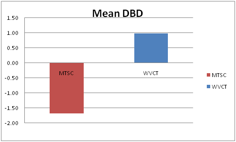
Matched-taper single-cone technique showed a higher overall difference in bone density (-1.67) than the warm vertical compaction technique (+0.99) (Figure 2). The minus sign indicates a decrease in bone density while a plus sign indicates an increase in bone density. However, this difference was statistically non-significant P > 0.05 at 95% level of confidence.

Figure 2 The mean difference in bone density (DBD) of matched-taper single-cone technique (MTSC) versus warm vertical compaction technique (WVCT).
Pain assessment
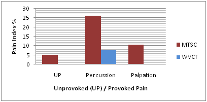
Unprovoked pain index:It revealed that warm vertical compaction technique recorded 0% after one year of treatment, while matched- taper single-cone technique recorded 5.3% (Figure 3).
Provoked pain index:Percussion test showed 7.8% for warm vertical compaction technique and 26.3% for matched-taper single-cone technique.
Palpation test showed 0% for warm vertical compaction technique and 10.5% for matched-taper single-cone technique (Figure 3).

Figure 3 Pain index percentage of unprovoked (UP) and provoked pain (Percussion and palpation) of matched-taper single-cone technique (MTSC) and warm vertical compaction technique (WVCT).
Although histological examination is considered as the most accurate standard to assess the health of the periapical tissues yet, the approach cannot ethically be applied in routine practice. Soon after its invention in 1895, radiographs had been used in the diagnosis of dental diseases. Radiographic imaging became a well-accepted surrogate measure for the histological condition of the periapex on the basis of a positive correlation between histological and radiographic findings.12
The sensitivity of the correlation between the histological condition of the periapical tissues and their radiographic appearance is variable among several studies. Sensitivity was recorded by Barthel et al.13 to be as low as 35%, while Brynolf12 and Green et al.14 recorded it to be as high as 88% and 66% respectively. Factors affecting sensitivity were all related to the relative mineral tissue loss including; the extent of the lesion,15 inflammation,16 thickness of the overlying cortical bone,17 and superimposition of the lesion by other anatomical structures.
Large cross-sectional studies from different countries have reported that the prevalence of apical periodontitis and other post-treatment periradicular diseases can exceed 30% of all root-filled teeth population.18-20 The outcome of endodontic therapy is generally assessed one year after treatment and is categorized as follows:
According to Jesslen et al. 24 the validity of a 1-year follow up is predictable in over 95% of the cases. The sensitivity of the human eye to the minor radiographic changes overtime is limited. A lot of unnoticeable changes could be missed. Hence, digital radiography helps to detect these minor changes. This study addresses long-term evaluation of the success of root canal treatment using the matched-taper single-cone obturation technique versus the warm vertical compaction technique. Post-treatment success indicators were both clinical and radiographic. One-year post-treatment subjective and objective evaluation of pain and assessment of radiographic bone healing in terms of periradicular bone density were the measures employed to verify success.
Limited information is available on the sealing quality of the new matched-taper single-cone root canal fillings as compared with that of gutta-percha warm vertical compaction. Although the use of dyes, radioisotopes, fluid filtration, bacteria, and endotoxin penetration techniques have been tried to evaluate the seal of endodontic materials,4 long-term assessment of periapical tissue reaction could be a good indicator.
In the current study, matched-taper single-cone technique had a similar long-term sealing ability to the warm vertical compaction technique as indicated by the absence of significant difference in bone density. This finding was consistent with Tasdemir et al.25 who found that filling with single-cone, lateral condensation, and warm vertical compaction techniques in canals treated with ProTaper or M two rotary instruments had similar levels of sealing efficacy.
The comparable sealing ability findings of both cold lateral and warm vertical compaction techniques could help to infer results of studies comparing the sealing ability of single-cone and cold lateral compaction techniques and the findings of this study.7 In agreement with our results, Gordon et al.,5 Romania et al.,26 Wu et al.,27 Inan et al.,28 and Marciano et al.9 found similarity in root canal filling quality of both single-cone and cold lateral compaction techniques.
In contrast to our findings, Schafer et al.7 found that warm vertical compaction technique produced significantly higher gutta-percha filled areas and lower sealer-filled areas than the matched-taper single-cone.
Upon clinical examination, most of the recorded unprovoked or provoked pain reactions were related to periodontal pocketing and food collection due to negligence of patients to restore their teeth with a proper final coronal restoration.
Within the limitation of this study, it seems that matched-taper single-cone obturation technique is an efficient and fast technique compared to warm vertical compaction technique. However, the role of a preceding efficient canal space cleaning and shaping is an important variable that must not be overlooked at any time.
The author declares that there was no conflict of interest.

©2015 Atteia, et al. This is an open access article distributed under the terms of the, which permits unrestricted use, distribution, and build upon your work non-commercially.