Journal of
eISSN: 2373-4345


Case Report Volume 9 Issue 2
1Oral & Maxillofacial Surgeon, Private Practice, Colombia
2Oral & Maxillofacial Resident Surgeon, University of Antioquia, Colombia
3OCB Clinic, Manizales, Colombia
4Dentist, Master in Laser-therapy, Clinica OCB, Manizales, Colombia
5Head for Education Swiss International Academy of Osseointegration, Switzerland
Correspondence: Jorge Ivan Cardona Estrada, Oral & Maxillofacial Surgeon, Private Practice, Pereira, Megacentro Pinares Torre 2 Office 809, Colombia, Tel 57 6 3212171
Received: March 01, 2018 | Published: March 6, 2018
Citation: Estrada JIC, Gómez NC, Herrera OCB, et al. Mandibular periosteal vertical distraction before dental implants placement: case report. J Dent Health Oral Disord Ther. 2018;9(2):108-113. DOI: 10.15406/jdhodt.2018.09.00339
Periosteal Distraction (PD) has been investigated in animals for several years with the purpose of vertically bone augmentations in atrophic areas where osteogenic distraction (OD) is not possible. However, up to date, PD has not been related in humans. A 63-year-old woman, with vertical and transverse atrophy of the left inferior alveolar ridge, was clinically and tomographically assessed. A custom made periosteal distraction device (Tracper 3) was placed under local anesthesia. It was activated 0.25 mm every 12 hours since the fourth day, for 15 days. Four months later, a 7.5 mm vertical bone increase was tomographically confirmed. The PD device was then removed and two dental implants were placed and loaded three months later. This clinical case clearly shows the potential of the PD technique in severe bone atrophy in humans. More extensive clinical studies are necessary to confirm the interest and precise clinical modalities of utilization of periosteal distraction in humans.
Keywords: periosteal distraction, alveolar jaw bone atrophy, bone regeneration, dental implants.
Undifferentiated periosteal mesenchymal cells subjected to mechanical stress, can be transformed into osteoblasts and produce bone neo-formation in a similar way to what happens in osteogenic distraction where an osteotomy is performed followed by progressive separation of the bone fragments that generate new bone formation.1,2 This technique is today recognized as being effective to increase the vertical dimension of edentulous alveolar crests with severe atrophy.3 Studies on PD in experimental animals for over 22 years have combined several elements to promote neo-osteogenesis: tricalcium phosphate,4 RTG membranes,5,6 lipid-lowering drugs,7 plasma rich fibrin PRF8 and stem cells9 among others. According to Zhao and colleagues, it is now possible to start the clinical investigation of this technique.10 We conducted a preliminary study in humans, applying a subperiosteal distraction device to increase the height of the edentulous alveolar ridge allowing to place implants in the newly formed bone.
A 63-year-old, non-smoker, female patient, with no relevant personal or family medical history, to whom two implants first right mandibular premolar and molar, had been placed six months previously, requested a similar treatment in the opposite side in the edentulous area distal to the first left mandibular premolar . The cone beam tomography (CB-CT) indicated a 10 mm alveolar height at level of the left second premolar with transversal deficiency and a 3.2 mm height at level the first left mandibular molar (Figure 1). The patient was informed of the necessity of a bone augmentation, with the possibility of utilization of heterologous or autologous bone graft or periosteal distraction, indicating her that this technique had only been performed in experimental animal studies and explaining her the risks and benefits of each option. The patient agreed the PD option and signed the informed consent, authorized by the Medical Ethics of Human Research Committee at CES University of Medellin – Colombia. After dental impressions, an acrylic model was produce to prepare the distraction device. The CB-CT area was 3D printed at DME3D in Medellin - Colombia, eliminating the cortex in front of the inferior dental nerve, in such a way that the position of the nerve was clearly indicated (Figure 2).
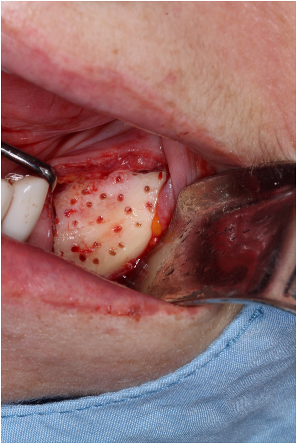
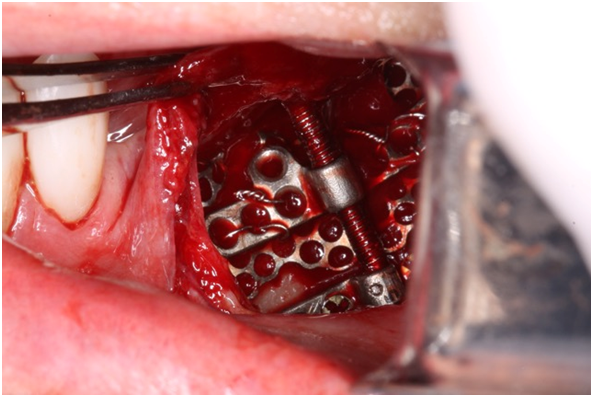
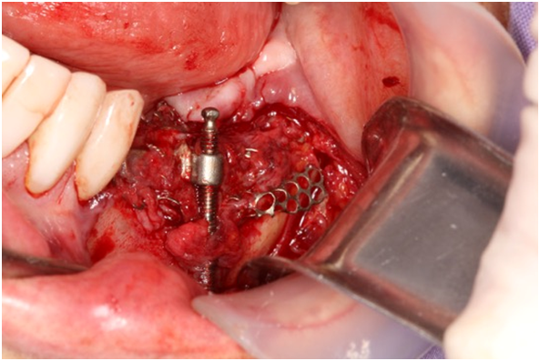
For the PD, a prototype device called TRACPER 3 was used. It was designed from an alveolar osteogenic distractor (Walter Lorenz , Jacksonville-Fl-USA), to which a 20 x 30 x 0.2 mm titanium mesh mm was fixed by means of a ligature wire, leaving it free to be adjusted and adaptated to the edentulous bone morphology. Under appropriate aseptic measures, the distraction device was placed under loco-regional anesthesia with 2% lidocaine and epinephrine. An incision of the mucoperiosteum at the level of the jugal sulcus of the 35 to 38 area was performed, followed by subperiosteal dissection until visualization of the basilar edge of the jaw, of the complete alveolar crest in the lingual side of the mandibular body and of the left mental nerve. Multiple perforations of the alveolar bone surface where performed and the periosteal distraction device was fixed with four 1.5 mm titanium screws to the mandibular base (Figure 3). The mucoperiosteum was then incised and finally sutured in two layers with Polyglyd 5-0 resorbable material., and a radiographic control was performed (Figure 4). Amoxicillin 500 mg (AmoxalR Glaxo Wellcome Mayenne –France ) 1 capsule every 8 hours for ten days and Nimesulide 100 mg (MesulidR Novamed S.A Barranquilla –Colombia) 1 tablets every 12 hours for five days were given.. Four days after placement the activation of the distraction device was initiated at a rate of 0.25 mm every 12 hours, after oral rinse during two minutes with 0.12% chlorhexidine digluconate (ClorhexolR Farpag Bogota – Colombia) . At the time of activation, the integrity of the oral mucosa and dental occlusion was checked (Figure 5). A small mesio-lingual dehiscence in the 35 area in relation with the anterior edge of the titanium mesh was observed on the fourth day of activation. To manage this dehiscence a Triticum Vulgare phyto-stimulant (Fitostimuline GelR Famaceutici Damor S.p.a Italy) was applied three times a day, the edge of the exposed mesh was cuted, and an acetate plate was placed to protect the left lateral side of the tongue. Distraction stage process was conducted on 15 days for a total of 30 activations, equivalent to 7.5 mm increase in alveolar height. Upon completion of the distraction, 12 daily sessions of intraoral 940 nm laser diode (Biolase , Cromwell, Irvine CA – US) , were applied with 0.2 W/continuous mode (CW). The hand piece was positionned at 8 mm from the mucosa moving the spot from mesial to distal of the PD area for 20 seconds on vestibular side and lingual side, in the goal to decrease the pain and inflammation of the ulceration that stay stable during the activation time. After the periosteal distraction, a CB-CT was performed to objective the result and to decide the length and diameter of the implants (Figure 6) (Figure 7). Removal of the periosteal distraction device and placement of the dental implants were carried out four months after the distraction. A fibrotic capsule was firmly attached to the device mesh (Figure 8). The clinicaly observed bone quality was favorable. The PD device was removed and two Bone Level implants NC Roxolid SLA ( Straumann, Basel Switzerland) were placed, 3.3 x 8 mm for 35 and 4.1 x 10 mm for 36 (Figure 9). A sample of bone tissue was harvested in 37 area , for histological analysis (Figure 10). Following the conventional loading protocol11 for osseointegration implant, ’ prosthetic rehabilitation was performed 3 months after implants placement (Figure 11) (Figure 12).


Figure 3 (A) Multiple perforation of the alveolar cortex; B: Placement of the custom made d istraction device whit fixation to the basal mandibular edge.

Figure 6 Tomography prior to removing the PD device. The most radiolucent zone corresponds to the neoformed bone on the preexisting alveolar ridge.

Figure 8 Removal of the distraction device, fibrotic capsule of the mesh, four months after the distraction process.
Several studies advise the indication of alveolar OD when loss of height is greater than 7 mm (4.12); however, in places where due to anatomical limitations or with very severe alveolar atrophy as in the case presented, it is not possible to perform safely an osteotomy, PD could become the most appropriate choice. Despite being a custom made device, TRACPER 3 fulfilled the stated objective, with no micro-movements, which could affect the formation of bone callus.13,14 The design of this prototype should be improved and may motivate to design osteoinductive devices,15 or self-regulated5 or to combine them at the time of placement with biological elements.8,9 The titanium mesh in narrow areas could be the origin of mucoperiosteum ulcerations during the distraction process, so the mesh should be carefully adjusted during placement of the distraction device, in order to avoid high pressure at the periphery and angles of the mesh on the mucoperiosteal flap. A specifically designed prefabricated mesh would be an avantage in this field. Perforation of the cortical bone prior to the placement of the distraction device and preservation of the integrity of the periosteum are proposed in the literature, to improve the results.13−16 Despite differences between animal and human models,17 the periods of latency (4 days), distraction (15 days at a rate of 0.25 mm every 12 hours) and maturation (4 months prior to dental implant placement) were adequate for human application of the PD technique. Several scientific articles have reported that laser application18 accelerates bone mineralization, so it would be of interest to conduct comparative studies of PD with/without laser utilization to precise the ideal clinical protocol. Following the results obtained in human clinical studies with OD,19−21 a three months delay before loading the dental implants after the PD process , allowed to succesfull osseointegration and resistance to 35 ncm torque at the abutment placement. The bone specimen harvested 4 months after the periosteal distraction process from the new bone formed in our patient features the histologic characteristics of natural lamellar bone, and therefore the conditions to place implants. Kessler et al.22 found no significant differences in the type and quantity of bone obtained through subperiosteal technique and autologous bone graft in calvarium bone of pigs. In opposition to Sencimen et al.,3 they stated that the bone obtained allows the placement of dental implants after a consolidation period of only six weeks.
Favorable preliminary results in this clinical case indicate that research carried out on animals has open the way to propose periostal distraction in case of severe atrophy in edentulous areas for future implant placements in patients. Further human controlled clinical studies would be necessary to confirm, the potential of periosteal distraction in atrophic bone and to precise clinical modalities and timing to allow succesfull results for implants placed in the gained bone.
Thanks to Doctor Maria Eugenia Becerra Herrera for her invaluable cooperation in the preparation of the paper.
Authors state having no conflicts of interest.

©2018 Estrada, et al. This is an open access article distributed under the terms of the, which permits unrestricted use, distribution, and build upon your work non-commercially.