Journal of
eISSN: 2373-4345


Case Report Volume 14 Issue 1
Department of Fixed Prosthodontics, University of Monastir, Tunisia
Correspondence: Imen Kalghoum, Department of Fixed Prosthodontics, Research Laboratory of Occlusodontics and Ceramic Prostheses LR16ES15, Faculty of Dental Medicine, University of Monastir, Monastir, Tunisia
Received: January 20, 2023 | Published: January 30, 2023
Citation: Jbeniany M, Hadyaoui D, El-Ayachi I, et al. Esthetic rehabilitation of maxillary incisors with porcelain veneer and lithium disilicate full coverage crown. J Dent Health Oral Disord Ther. 2023;14(1):4-8. DOI: 10.15406/jdhodt.2023.14.00586
The aim of this article is to show the interest of minimally invasive restorations (ceramic veneers and lithium disilicate full coverage crown) to restore the appearance of discoloured, fractured teeth or teeth with diastemas providing esthetic results and solving problems related to the size, shape, and color of the anterior upper teeth.
For this purpose, a correct diagnosis, careful treatment plan, clinical and laboratory procedures offered a satisfying esthetic outcome. Moreover, digital design can be used to enhance the final result since it offers a better predictability of the proposed treatment and facilitates communication with patient. It can be concluded that lithium disilicate restoration is a predictable and long lasting treatment option with which not only esthetics but also the strength and function of teeth can be re-established. The objective of this case report is to highlight the steps in dental rehabilitation using ceramic veneers reinforced by lithium disilicate. The objective of this case report is to describe all steps of dental rehabilitation using ceramic restorations reinforced by lithium disilicate.
Keywords: porcelain veneers, lithium disilicate, full coverage crown, esthetic restoration, smile design
The innovation and development in restorative materials, bonding and ceramics offer, nowadays, to clinicians a whole plethora of esthetic treatments to preserve a natural appearance while respecting the therapeutic gradient based on tooth tissue economy.1–4
Since their introduction in 1983, ceramic veneers have been considered as one of the most viable treatment modalities not only for their esthetic outcome and minimal thickness but also for their strength, longevity and biocompatibility.5
Indeed, ceramic veneers establish esthetics, function for discoloured, fractured, malposed or malformed teeth. They can also be performed for diastema closure. Such esthetic treatments, however, must not be conducted without an appropriate restorative planning using digital design or wax-up. This concept of planning offers not only a better predictability of the proposed treatment but also a guided preparation to minimalize dental removing. When it comes to preparations, they concern the buccal surface with a proximal finish and cervical preparation. The incisal edge can be prepared or not, depending on the preparation category: overlap or non-overlap.
Overlap prepared veneers are mostly indicated in cases of third incisal fracture. It reinforces the incisal edge and offers a better esthetic appearance enabling good light propagation in the third incisal. In some cases of slightly lingual tipping teeth, incisal overlap may help to reposition the tooth.
However, in case of endodontically treated anterior teeth varying treatment protocols and methods of preparation have been indicated depending on crown length and type of material. Bonded porcelain crown seems to require a minimally invasive preparation and offer an esthetic result for those non-vital teeth.
Hence the aim of this paper is to describe the possibility of esthetic re-estabilising with a minimal invasive restoration according to vital and not vital teeth.
Case history, diagnosis
A 22-year-old girl had been referred to the Department of fixed Prosthodontics in dental university of Monastir, Tunisia.
She was complaining about a discoloured composite restoration on teeth number 11 and 21(Figure 1). She wanted to improve their shape and color. She was also asking for a small adjustment of the alignment of the whole maxillary anterior group of teeth.
After anamnesis and clinical examination, we reported a double oblique fracture line on tooth number 21 exceeding the incisal third. The composite restoration was extensive and surrounded by a large dyschromia. The fracture line on tooth number 11 was only defined on the incisal third. Gingival contour was compromised with a red and puffy papilla (Figure 2). The patient was very anxious about her unpleasant smile.
Periapical radiograph did not reveal any periapical pathological condition but an unsatisfactory endodontic treatment (Figure 4).
In order to obtain better predictability of the treatment, dental casts were manufactured. In addition, we conducted extra and intraoral photographs that allowed a digital plan thanks to DSD technique.
Whereas digital planning required three photographs as follow: face with a wide smile (Figure 1), intraoral view of the full maxillary arch with teeth before treatment (Figure 2) and the "12 O'clock" view allowing the buccal projection of incisal margins to be assessed in relation with the position of the curve line of lower lip (Figure 3).
Digital analysis of initial patient’s smile reported a flat curve of anterior teeth which is not parallel to the lower lip line. The dental midline was shifted mesially due to composite restoration on tooth 21 (Figure 5). The maxillary arch photography allows the analysis of ideal proportions of maxillary incisor teeth (Figure 6).
Standard template of maxillary incisor was used to preview the bi-dimensional final result including esthetic correction of incisor line and shape. Esthetic enhancement of the gingival contours should begin with new proposed tracings (Figure 7 & 8).
Mock-up and dental preparation
Diagnostic wax-up was preformed based on digital smile design then it was duplicated on patient’s teeth through a silicone guide filled with bis-acryl resin to perform an intra-oral esthetic mock-up simulating the final result in a reversible phase.6–8
Mock-up does not only enable a better visualization of the final prosthetic project for the patient but also is used as a template to create a sufficient space for restorative material while preserving maximal dental enamel. After validation of the final esthetic result, an overlap veneer on tooth number 11 was indicated where as tooth number 21 required a full crown vitro-ceramic restoration.
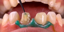
We started the preparation of tooth number 11 guided by the diagnostic mock-up. The tooth preparation was restricted entirely to the enamel uniform and well rounded. In this case, the recommended thickness of the porcelain veneer is 0.7 mm on the buccal surface followed with a reduction of 1.5mm for the incisal edge and a 0.5 mm palatal chamfer in the middle third.
Initially, horizontal slots in the region of the middle and cervical third with the depth marker-bur and two incisal grooves with a round ball-tip diamond bur were performed. Retraction cord was inserted and the preparation was completed with a round-end tapered diamond bur. The polishing of the preparation was done with sequential disks and polishing rubber points. Finally, all angles were rounded.
For the endodontically treated tooth a bonded porcelain crown was indicated. Tooth architectural preparation started by removing 2 mm in the incisal edge followed by a 1.0 mm axial margin width using a rounded tapered diamond bur. Porcelain crowns require a 10 degrees angle as a total occlusal convergence followed by polishing and rounding all angles (Figures 9-11).
Temporisation
The temporary veneer and crown were made using a bis-acryl temporary resin and were cemented with non-eugenol cement to preserve enamel conditioning for bonding.
Temporary restorations allow to evaluate the correct aesthetic and occlusal positioning of anterior maxillary teeth showing the relationship between the curve of the incisal edge of the maxillary anterior teeth and the curve of the upper edge of the lower lip. They should be harmonious during voluntary smile and should respect the golden standard of dental proportions and incisal edge position9 (Figure 12).
Bonding
Prior to cementing, the porcelain restorations were carefully positioned with try-in pastes (RelyX Veneer try-in-paste) to verify marginal adaptation, alignment, shape, and color. (Figures 13 & 14)
For luting, the conditioning of the internal surfaces of the restorations was treated with alcohol solution. A layer of 9% hydrofluoric acid was then applied for 20 seconds (Ceram-etch 9% Itena); it was then washed with water and air-dried. A silane (Silan-it Itena) followed by adhesive (3M ESPE Single Bond Universal Adhesive Bonding Agent) were then applied (Figures 15–20).
Tooth surface (enamel and dentin) treatment
A light cured dam and a retraction cord were established for isolation. The teeth were cleaned with pumice and water, etched with 37% phosphoric acid for 30s (Scotchbond universal etchant 3M), washed, and dried.
A simplified universal adhesive system (3M ESPE Single Bond Universal Adhesive Bonding Agent) was applied with photo activation. The light-cured resin cement was applied first, to the veneer which was carefully positioned on the preparation then to the inner surface of the crown after that both were light cured for 3 seconds. The latter allowed cement excess removal with a brush and a dental floss, finally the procedure was followed by light curing for 40 s from palatal, labial, and incisal sides. Excess luting cement was removed and the marginal area was finished and polished with abrasive discs and strips. Occlusal contacts were marked, and protrusive and lateral movements were checked. The patient was satisfied with her new smile line and excellent view of the anterior teeth (Figures 21–25).

Figure 24 Dental surface treatment with universal adhesive system (3M ESPE Single Bond Universal Adhesive Bonding Agent).
A simplified universal adhesive system (3M ESPE Single Bond Universal Adhesive Bonding Agent) was applied with photo activation. The light-cured resin cement was applied first, to the veneer which was carefully positioned on the preparation then to the inner surface of the crown after that both were light cured for 3 seconds. The latter allowed cement excess removal with a brush and a dental floss, finally the procedure was followed by light curing for 40s from palatal, labial, and incisal sides. Excess luting cement was removed and the marginal area was finished and polished with abrasive discs and strips. Occlusal contacts were marked, and protrusive and lateral movements were checked. The patient was satisfied with her new smile line and excellent view of the anterior teeth (Figures 26–29).
Preparations for ceramic veneers were described according to enamel preservation varying from minimum preparation maintaining approximately 95% of enamel to tooth reduction up to 1.0 mm and should allow, whenever possible, a reservation of at least 50% of enamel10 to ensure chemical adhesion and clinical longevity.7 A longitudinal study with a 12-year follow-up has shown that ceramic veneers cemented on enamel have higher clinical longevity than those cemented on dentin, with success rates of 98.7% and 68.1%, respectively.11
In case of palatal extension, the preparation should never be limited on the area of mechanical constraints such as the concavity just below the cingulum and occlusal contacts must be placed on either ceramic surface or tooth surface but never on the junction in between.
Overlap veneer offers better esthetic appearance, good light propagation in the incisal third and help to reposition the tooth. Additionaly, as it has a single path of insertion it prevents any displacement of the veneer during cementation.10,12
Many ceramic systems can be used to restore anterior teeth; their indication depends on tooth preparation design, minimal thickness of ceramic corresponding to mechanical strength and esthetic result. Lituim discilicate ceramic is the most frequently indicated to restore anterior teeth due to its natural appearance, translucency and mechanical strength with 1 mm minimal thickness.12 Moreover, their chemical nature allows inner surface etching with hydrofluoric acid associated with the use of silane coupling agents offer a long term longevity of the tooth ceramic bonding interface.
Before bonding, evaluation of the marginal adaptation, shape and color should be checked. It is recommended to use the try in paste with similar color shade, of the final resin bonding, to simulate the final esthetic result. They also allow an easier placement of veneers during the clinical trial.
The success of the porcelain veneer is greatly determined by the strength and durability of the bond interface formed between the three different components: tooth surface, porcelain veneer, and luting composite.
The type of the resin cement and its long term color stability are important factors to achieve aesthetic success; especially in case of highly translucent porcelain laminate veneers.
The dual-polymerizing cement used for bonding ceramic veneers to enamel has higher color variation than the light-polymerized materials which have been shown to possess bond strength and higher colour stability.13
Minimally invasive restoration approach using lithium disilicate and conducted by a careful planification allows to restore natural mimmicry and teeth harmony with a smile curve parallel to lower lip line. Esthetic and functional results are offered thanks to optical properties of both ceramic and bonding materials.
None.
The authors have no conflict of interest.

©2023 Jbeniany, et al. This is an open access article distributed under the terms of the, which permits unrestricted use, distribution, and build upon your work non-commercially.