Journal of
eISSN: 2373-4345


Case Report Volume 15 Issue 3
1Professor and Head of Oral and Maxillofacial Surgery Department, Al-Azhar University, Egypt
2Researcher PhD of Oral and Maxillofacial Surgery, Faculty of Dental Medicine, Al-Azhar University, Egypt
Correspondence: Prof Ayman Hegab, Professor and Head of Oral and Maxillofacial Surgery Department, Faculty of Dental Medicine, Al-Azhar University, Egypt
Received: June 29, 2024 | Published: August 19, 2024
Citation: Hegab A, Elmasry M. Conservative management of giant follicular ameloblastoma: report of two cases. J Dent Health Oral Disord Ther. 2024;15(3):146-149. DOI: 10.15406/jdhodt.2024.15.00627
The treatment of choice of Ameloblastoma is surgical resection which resulting in large jaw defects. The challenge in managing mandibular giant ameloblastoma is in achieving complete excision and reconstruction of the defect. However, conservative non-surgical approach was mentioned in the literature as a useful approach in young patients. The present article reported two cases of conservative treatment of mandibular giant follicular ameloblastoma in young patient to decrease the tumour size and subsequently the resected segment of the mandible and in another inoperable case due to her medical problems. Good results could be achieved in the treatment of large Giant Ameloblastoma using marsupialization in the modern era of more aggressive surgical procedures and reconstructive techniques. Marsupialization revealed to be more advantageous in many respects and is therefore considered a worthwhile procedure to reduce the tumor size before resection and can be used in inoperable cases.
Keywords: ameloblastoma, conservative treatment, carnoy’s solution
Ameloblastoma is a benign odontogenic tumor of the jaws with locally aggressive behavior and a high recurrent rate. Ameloblastoma is the most common odontogenic jaw tumour, accounting for 1% of all cysts and tumours of the jaw and 11% of all odontogenic tumours.1 In general, ameloblastoma can be categorized into 3 types: solid multicystic, unicystic and peripheral ameloblastoma. Solid Ameloblastoma is a slow growing, but locally invasive, tumor. Some ameloblastomas become gigantic and destroy adjacent tissues. Conversely, unicystic ameloblastoma possesses a less aggressive nature and a lower recurrent rate. It can be either a tumor de novo or a tumor arising from an odontogenic cyst. Both solid and unicystic ameloblastoma commonly occur in the mandible, especially the molar-ramus area.2–4
Various treatment modalities for ameloblastoma have been reported. However, the universally accepted approach remains unsettled. It can range from conservative treatments, such as enucleation with or without curettage, to aggressive treatments which include peripheral osteotomy and resection.1,2,5 Once mandibular ameloblastoma becomes gigantic, it requires a radical approach.2,3 Some authors reported the treatment of extensive ameloblastoma using radical excision with immediate Micro vascular reconstruction.6 Whereas R. Madhup et al successfully treated a giant mandibular ameloblastoma by radiotherapy.7
In spite of these many treatment methods identified in the literature, there is still controversy about the therapy based on the clinical presentation or histopathological characteristics of the tumors. The description of definite treatment of giant mandibular ameloblastoma still controversial. This is a report of conservative management of two cases of mandibular giant follicular ameloblastoma.
Case 1
A 21-year-old female patient referred to our oral & maxillofacial surgery department with chief complaints of a slowly growing swelling on the right side of the face for two years. The swelling involve the parotid region with facial asymmetry. There was no associated pain, difficulty in opening the mouth, chewing or articulating. On physical examination, there was a hard non-tender mass arising from the right side of the mandible, involving the ramus and angle of the mandible. The oral mucosa was normal. No neck nodes were palpable. Systemic examination was normal. An orthopantomogram (OPG) showed large multilocular radiolucent lesion in the right side of mandible involving the mandibular angle, ramus, coronoid process and mandibular condyle and associated with impacted third molar tooth (Figure 1A). CT scan showed that the lesion was confined to the mandible, with a thinned out cortex. No cortical bone destruction or soft tissue invasion of the tumour mass was noticed (Figure 1B). Incisional biopsy was taken in the outpatient clinic under local anaesthesia and the histopathologic examination confirmed follicular ameloblastoma (Figure 1C). The primary surgical plan was to make hemi mandibulectomy. Nonetheless, we changed this plan and decided to make enucleation, curettage and chemical cattery by carnoy’s solution first to decrease the tumour size and respectively the resected segment of the mandible. Under general anaesthesia, the patient treated by enucleation, curettage with extraction of the impacted third molar and second molar teeth followed by chemical cattery by carnoy’s solution (chloroform 3 ml, absolute alcohol 6ml, glacial acetic acid 1ml, ferric chloride 1g). The carnoy’s solution was applied into the bone after complete enucleation and curettage and allowed to remain for 5 min. The bone cavity was soaked with the carnoy’s solution for another 5 min.
The patient was reviewed postoperatively in the outpatient clinic every 2 months in the first year and then every 6 months. Clinical and radiographic examination of the patient revealed complete healing of the lesion with no recurrence up to 7 years postoperatively (Figure 1D).
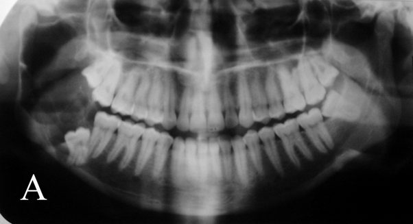
Figure 1a Orthopantomogram showed large multilocular radiolucent lesion in the right side of mandible involving the mandibular angle, ramus, coronoid process and mandibular condyle and associated with impacted third molar tooth.
Case 2
A 78Y female patient came to the outpatient clinic with huge neglected mandibular swelling in the right side. The patient provided history of painless swelling started from about 7Y and gradually increase in size. The swelling caused loosing of the associated teeth and resulted in extraction of the teeth. The patient sought medical care only after the swelling became associated with severe pain. The patient contributed past medical history of uncontrolled hypertension, uncontrolled diabetes, bronchial asthma and heart failure. The woman had been evaluated 6 years earlier for an enlarging jaw mass, and a biopsy at that time revealed an ameloblastoma. She had been offered surgical excision, but the surgery was cancelled due to her medical condition. Clinical examination showed firm huge swelling extend from the Symphysis region to the mandibular angle with severe facial asymmetry. The swelling extend to the submental and submandibular regions (Figure 2A). Intraoral examination showed huge swelling extends to the sublingual region with obliteration of the buccal vestibule (Figure 2B). Due to the tumour size that cannot be evaluated by the OPG and the medical condition of the patient that prevent radiographic evaluation by CT, the only available x-rays were Postro-anterior and lateral mandibular x-rays. Radiographic evaluation revealed multilocular radiolucent lesion with sever destruction and thinning of the inferior border of the mandible (Figure 3A & B). Aspiration of 60 cc of cystic fluid followed by incisional biopsy was performed in the outpatient clinic under plain local anaesthesia. The histopathologic examination confirmed follicular ameloblastoma (Figure 3C). The treatment plan was made on the basis of the medical condition of the patient. Under plain local anaesthesia in the outpatient clinic, intraoral decompression of the tumour with suturing of the lining to the oral mucosa was performed. One week later, impression was taken and construction of acrylic stent with central hole to maintain the opening was done. 6 months later, the patient showed noticed decrease of the tumor size with disappearance of the pain (Figure 4A). Two years postoperatively, Radiographic evaluation by OPG revealed significant reduction of the tumor size with formation of good amount of new bone at the inferior border of the mandible preserving the mandible from the pathologic fracture (Figure 4B).
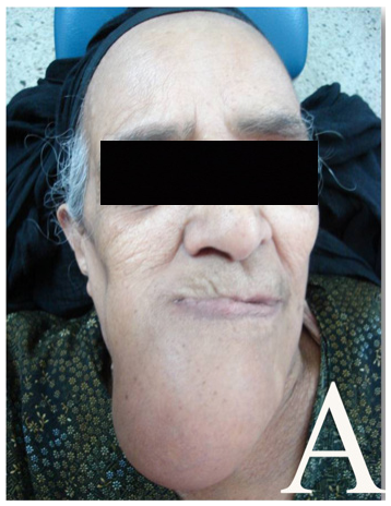
Figure 2a Extra oral view showed giant swelling extend from the Symphysis region to the mandibular angle with severe facial asymmetry.
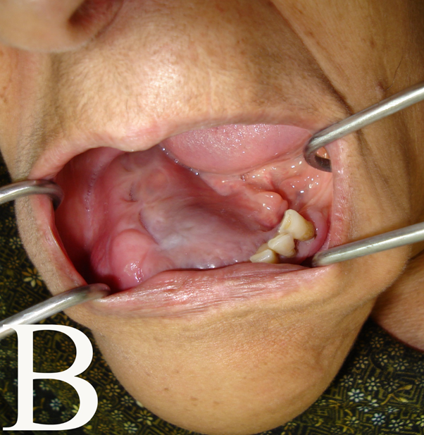
Figure 2b Intraoral view showed huge swelling extends to the sublingual region with obliteration of the buccal vestibule.
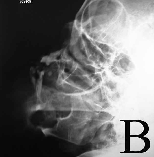
Figure 3b Lateral mandibular x-rays. Radiographic evaluation revealed multilocular radiolucent lesion with sever destruction and thinning of the inferior border of the mandible.
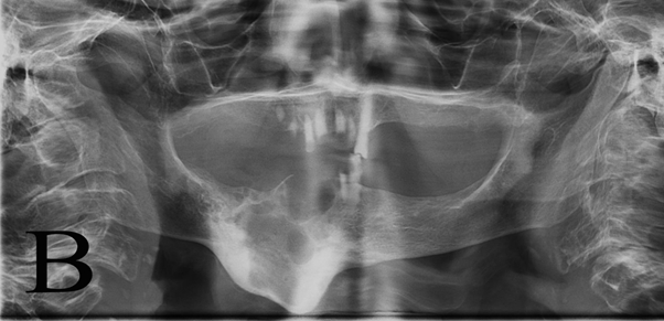
Figure 4b Orthopantomogram revealed significant reduction of the tumor size with formation of good amount of new bone at the inferior border of the mandible.
Recurrent infection that required regular irrigation through the acrylic stent with the antibiotic coverage depending on the culture sensitivity test is the only complication that occurred with the patient.
Ameloblastoma is a benign, but locally aggressive, odontogenic tumour. A conservative approach usually yields a high recurrent rate, thereby requiring close vigilance. Repeat treatment of a small recurrence is more acceptable than jaw amputation with complex reconstructive surgery. An extensive tumour may be destructive and life-threatening, necessitating adequate excision which depends upon its site and extension. Until now, there has been neither definition of nor definite treatment consensus on giant mandibular ameloblastoma. Current opinion regarding treatment of ameloblastomas is essentially based on case reports, anecdotal evidence, retrospective reviews, and histological evidence. There are not many large-scale studies with long-term follow-up results.8 The benign nature of these lesions often leads the surgeons to perform simpler extirpative procedures to avoid the potential morbidity associated with large resections. This approach is still commonly practiced, despite reported recurrence rates of 55% to 90% for solid multicystic treated by enucleation or curettage and even occasional metastases.3,8,9
Late presentation of ameloblastoma usually involved a large part of the mandible. Surgical treatment at this stage will result in hemi mandibulectomy, which involves the resection of the bone to which the musculature of the floor of the mouth and the tongue are attached. Serious aesthetic and functional problems are encountered, necessitating the reconstruction of the lost tissues. There is a serious reduction in the quality of life of these patients regarding feeding, speech, appearance and saliva control as a result of lack of lip support.
Treatments such as enucleation and peripheral ostectomy or physicochemical treatments including liquid nitrogen or Carnoy’s solution have been suggested; initial studies are promising but long-term results are not available.10
In the present case report, conservative treatment was completely successful in the case treated by enucleation, curettage and chemical cattery by carnoy’s solution. The rationale for using carnoy’s solution was that it can penetrate the cancellous bone and fix and devitalize the cells may remain in the jaw bone after enucleation and curettage of the ameloblastoma. The treatment plan was to use enucleation and curettage to decrease the tumour size and the resected bone segment, but the result was complete healing with no recurrence up to 7 years postoperatively. In the second case, the medical condition of the patient prevented the use of radical approach (inoperable). Conservative treatment resulted in disappeared of the pain, significant reduction of the tumour size with new bone formation at the inferior border that preserved the mandible from pathologic fracture.
These cases further highlight the role of conservative treatment of advanced, giant mandibular ameloblastoma. It can be used as first stage treatment to reduce the tumor size before resection and can be used in inoperable cases. Nevertheless, our report seems not to be a conclusive assessment. A larger number of cases and longer follow-op periods remain necessary.
None.
The authors declare that there are no conflicts of interest.

©2024 Hegab, et al. This is an open access article distributed under the terms of the, which permits unrestricted use, distribution, and build upon your work non-commercially.