Journal of
eISSN: 2373-4345


Case Report Volume 9 Issue 2
1Department of Dentistry, University of S?o Paulo School of Dentistry, Brazil
2Department of Dentistry, CESUPA School of Dentistry, Brazil
3Department of Stomatology, University of S?o Paulo School of Dentistry, Brazil
Correspondence: Solange Kobayashi-Velasco, Department of Dentistry, University of S?o Paulo School of Dentistry, Brazil, Tel 55 11 96180-6060
Received: October 28, 2017 | Published: March 6, 2018
Citation: Kobayashi-Velasco S, Santos RLO, Salineiro FCS, et al. Comparison between panoramic radiography and CBCT imaging on the diagnosis of second molar external root resorption associated with an impacted mandibular third molar: two case reports. J Dent Health Oral Disord Ther. 2018;9(2):115-117. DOI: 10.15406/jdhodt.2018.09.00340
Third molar impaction is a usual condition found at the dental practice. It may be associated with external root resorption (ERR) of the adjacent second molar. In those cases, it is necessary to extract the third molar, so that it does not damage the second molar root. Periapical and panoramic radiographies may be employed to evaluate the third molar condition. Nowadays, cone beam computed tomography (CBCT) imaging is also used to analyze third molars. The aim of this paper was to report two cases of mandibular impacted third molar associated with second molar external root resorption, comparing panoramic radiography and CBCT imaging. Case report 1 described a panoramic radiography (PR) with superimposition between teeth 47 and 48, suggesting ERR, which was confirmed by CBCT imaging. Case report 2 depicted a PR that showed overlapping of teeth 37 and 38, suggesting ERR. CBCT imaging reported a discreet root flattening at 37, while 38 was located at the lingual aspect of 37. The position, inclination and location of the third molar may influence the location and severity of the ERR. CBCT exams are highly relevant for the analysis of impacted mandibular third molar associated with second molar external root resorption, thus consisting in an important tool for the practitioner during the planning, treatment and prognosis of each patient.
Keywords: Molar, third, Root resorption, Radiography, panoramic, Cone beam computed tomography
Third molar impaction is a usual condition found at the dental practice. Occasionally, it is necessary to remove the tooth in order to avoid other problems such as pericoronitis, swelling, odontogenic cysts or tumors, bone loss and external root resorption (ERR) of the adjacent second molar.1 However, the decision of extractiong or maintaining a third molar is controversial.2 If an impacted third molar is not extracted after a certain stage of its formation, it may contribute to second molar ERR.1 According to Tsesis et al.3 ERR is a pathological process that occurs at the permanent tooth outer surface and it may be induced by pulpal infection or periodontal inflammation related causes, or pressure associated with orthodontic movements, impacted tooth or pathoses. Periapical and panoramic radiographies (PR) are the standard imaging modalities for determining third molar characteristics. These imaging exams are readily available and dental professionals are acquainted with images interpretation. However, they are two-dimensional images, thus impairing a more detailed analysis of the region. Additionally, panoramic radiographies (PR) present a certain degree of distortion.4 The limitation generated by the absence of a third dimension resulted in methods that do not inform precisely the actual location of the third molar, its anatomy or its relationship with adjacent structures.4 Cone beam computed tomography (CBCT) imaging allows a tridimensional evaluation of teeth and their adjacent anatomical structures,4 resulting in a detailed visualization of the third molar as well as its neighboring structures, and subsequently, a meticulous diagnosis of ERR. CBCT is widely recommended for third molar surgical planning.4 Among the advantages of this imaging modality, some authors mention spatial resolution, multiplanar reconstruction simultaneous analyses and structures superimposition avoidance.5 Hence, the aim of this study was to report two cases of mandibular impacted third molar associated with second molar external root resorption, comparing panoramic radiography and CBCT imaging.
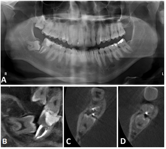
Case 1: A 24 year-old Caucasian male patient was submitted to a follow-up PR nine months after the extraction of tooth 38. The radiologist observed a severely inclined, mesialized and impacted 48. Its roots were contiguous to the mandibular canal roof and it was possible to recognize a severe dilaceration at the mesial root apex. Tooth 47 was endodontically treated and an overlap was noted between 48 crown and dental follicle, and 47 distal root (Figure 1A). Tooth 48 was recommended for extraction and a CBCT was requested for planning the surgical intervention. The patient was submitted to a CBCT exam with the following characteristics: field of view (FOV) 5.0 cm by 5.5 cm, voxel 0.15 mm, 96 kV and 12 mA (Planmeca ProMax 3D, Planmeca Oy, Helsinki, Finland). CBCT images revealed a severe ERR at tooth 47 distal root with the preservation of the endodontic filling material. Tooth 47 ERR was associated to tooth 48 crown (Figure 1B); the third molar crown was partially filling the space of the second molar distal root. The mandibular canal cortical was adjacent to 48 both roots (Figure 1B) (Figure 1C) (Figure 1D).

Figure 1 Case 1. A: Panoramic radiograph; B: CBCT exam, sagittal image, root resorption was induced by element 48 crown; C & D: CBCT exam, axial images, the mandibular canal cortical was adjacent to element 48 roots and the tooth 47 preserving only the endodontic filling material.
Case 2: A 31 year-old Caucasian female patient reported pain at the left retromolar trigone region. After clinical evaluation, the patient was submitted to a PR, and a CBCT exam for surgical planning (extraction of element 38). In the panoramic image, tooth 38 was in vertical position, with both mesial and distal roots overlapping tooth 37 distal root (Figure 2A). Apical portions of tooth 38 were covering the mandibular canal. A CBCT exam was performed at Vatech PaX-i3D (Vatech Co., Hwaseong-si, South Korea) device using the following parameters: FOV 5.0 cm by 5.0 cm, voxel 0.12 mm, 90 kVp and 5 mA. The images revealed that teeth 37 and 38 roots were contiguous (Figure 2B) and a discreet flattening of 37 distal root, possibly caused by the third molar roots mechanical pressure, was observed. Tooth 38 apical portion was neighboring the mandibular canal roof (Figure 2C) (Figure 2D).
A thorough anamnesis and clinical examination must always be performed before planning a third molar removal surgery. The information collected on these procedures will contribute for appropriate choices towards requesting proper imaging exams for each case.6 Oenning et al.7 compared PR and CBCT imaging of third molars and found out that 22.88% of external root resorption were diagnosed through CBCT exams, while only 5.31% were observed in PR. This study did not correlate a third molar particular position with a propensity of developing ERR on the second molar. However, according to our findings, we suggested that the position, inclination and location of the third molar may influence the severity of second molar ERR, which is in agreement with Matzen et al.8 and Oenning et al.9 studies. The third molars analyzed in our cases were either mesially inclined (Case 1) or in vertical position (Case 2). The same studies suggested that a CBCT exam obtained after the PR, might determine an adjacency between the second molar and the impacted third molar, corroborating the findings observed at our cases.
In Case 1, it was possible to observe, by using PR, an overlapping between tooth 47 distal roots and tooth 48 dental follicles, strongly suggesting an image of ERR. However, in the CBCT imaging, ERR was clearly detected, and then it was possible to observe a mesially inclined third molar that caused severe ERR at tooth 37 distal aspect of the distal root. Case 2 presented a superimposition between tooth 37 distal root and tooth 38 mesial root, suggesting an ERR at the panoramic radiography, while CBCT images confirmed only a discreet flattening at 37 distal root. Both third molar roots were observed at a distal-lingual relationship with the second molar, resulting in a flattening of the lingual aspect at tooth 37 distal root. This flattening might lead to a more severe ERR in the future, thus recommending the extraction of 38. PR from both our cases showed superimposed images of the second and third molars, hence not categorically confirming or rejecting the presence of second molar ERR. Besides providing the confirmation of ERR, CBCT imaging from both our cases determined a more precise view of the anatomical characteristics of the third molar, as well as its positioning in relation to the second molar and adjacent structures such as inferior alveolar nerve and buccal, lingual and mandibular base cortices.6-11 These images aided in planning and executing the surgical procedures. CBCT imaging are highly relevant for the analysis of impacted mandibular third molar associated with second molar external root resorption, thus consisting in an important tool for the practitioner during the treatment planning, and surgical outcomes.
Funding provided by CNPq - National Counsel of Technological and Scientific Development (SKV, PhD scholarships), and CAPES - Coordination for the Improvement of Higher Education Personnel (RLOS and FCCS, PhD scholarships).
The authors declare no personal or ethical conflicts of interest.

©2018 Kobayashi-Velasco, et al. This is an open access article distributed under the terms of the, which permits unrestricted use, distribution, and build upon your work non-commercially.