Journal of
eISSN: 2373-4345


Case Report Volume 15 Issue 4
1Department of Multidisciplinary Health, University Center Mauricio de Nassau (UNINASSAU), Brazil
2Department of Dentistry, Universidad Federal de Juiz de Fora UFJF, Brazil
3Post Graduate Student, Ph.D. Program, Department of Dentistry, Pontifícia Universidad Católica de Minas Gerais, Brazil
4Professor of Maxillofacial Surgery, Department of Dentistry, Universidad Estadual de Montes Claros UNIMONTES, Brazil
5Department of Restorative Dentistry & Prosthodontics, the University of Texas Health Science Center at Houston (UT Health) School of Dentistry, USA
6Postgraduate Student - Master Degree Program, Craniofacial and Cleft Lip and Palate at the Hospital for Rehabilitation in Craniofacial Anomalies of the University of São Paulo (HRACUSP), Brazil
7Department of Oral and Maxillofacial Surgery, Universidad Federal de Juiz de Fora UFJF, Brazil
Correspondence: Jefferson Matos, Department of Multidisciplinary Health, University Center Mauricio de Nassau (UNINASSAU), Juazeiro do Norte - CE, Brazil
Received: September 25, 2024 | Published: October 18, 2024
Citation: Matos JDM, Lima GO, Rodrigues ML, et al. Bilateral maxillary odontogenic fibroma: a case of rare occurrence. J Dent Health Oral Disord Ther. 2024;15(4):160-163. DOI: 10.15406/jdhodt.2024.15.00630
Aim: To report the case of a patient diagnosed with bilateral odontogenic fibroma in the maxilla that was surgically removed.
Clinical conduct: A 32-year-old female patient was referred to the Santa Casa de Montes Claros Hospital for the removal of a lesion in the maxilla. Intraoral examination revealed a mixed lesion in the maxilla, bilaterally, with a suspected odontogenic tumor. Cone beam computed tomography showed an expansive lesion in the posterior region of the right and left maxilla, extending to the palatine and zygomatic processes. An incisional biopsy was performed and revealed the proliferation of spindle cells amid collagenized stroma, with the presence of mitoses and foci of concentric calcification, accompanied by lacerated bone trabeculae and fibroconnective tissue with edema, which were compatible with the diagnosis of odontogenic fibroma. During removal, the lesion detached easily and it was necessary to remove a premolar corresponding to the area involved, which presented advanced mobility due to loss of support, followed by adequate cleaning of the surgical site.
Results: After the surgery to remove the lesion, the patient remained under clinical follow-up to evaluate the evolution of the case as well as its possible complications. Conclusion: This case highlights the importance of a careful clinical, imaging, and histopathological approach to lesions in the maxilla, for correct diagnosis and referral of the patient for appropriate treatment.
Keywords: surgery, oral, CBCT, incidental finding, oral and maxillofacial region.
Central odontogenic fibroma (COF) is a rare type of benign tumor of odontogenic origin that affects the gnathic bones, with an incidence ranging from 0% to 5% among all other odontogenic tumors.1 They are defined by the World Health Organization (WHO) as ectomesenchymal tumors originating from remnants of the periodontal ligament, dental follicle or dental lamina2,3 and that present quantitative variation of odontogenic epithelium amidst the mature fibrous connective tissue stroma.4
A COF can affect both the mandible and the maxilla in the same proportion of cases. When it occurs in the maxilla, it usually affects the anterior region. In the mandible, the premolar and molar regions are more affected.4–6 There is a slight predilection for females in a proportion of approximately 3:1 when compared to males.2,3,7,8 The average age of onset is 40 years, but there are reports in the literature of patients aged 4 to 80 years diagnosed with COF.1,3,8
Radiographically, the COF presents as a radiolucent image, well delimited and circumscribed by a sclerotic halo, and may be uni or multilocular, with displacement and tooth resorption.2,3,7,8,12–14 When there is the expansion of the cortical bone6,9,16 and tooth displacement, there is a high possibility of it being associated with the crown of an impacted tooth.4,9,10
From a histological point of view, there is a subdivision according to the amount of inactive epithelium found in the middle of the stroma, being: simple type (poor in epithelium) and WHO/complex type (rich in epithelium).4 The first is characterized by fine collagen fibers, considerable amorphous ground substance, and stellate fibroblasts organized in a spiral pattern.4,10 Calcifications may be present due to remnants of the inactive odontogenic epithelium.4,6,10,19,20 In the WHO type, long cords or isolated nests of inactive epithelium interspersed with numerous small blood vessels are observed throughout the lesion.4,6,10,19 There is variation in the fibrous component, which may range from myxoid to hyalinize. When calcifications are present, it is a collagen matrix remaining from a tubular dentin, osteoid cementum, or dysplastic.1,8,14,17 Conservative surgical treatment by enucleation followed by curettage is the most recommended, with high success rates, low recurrence, and low potential for malignant transformation.2,3,7,11–15
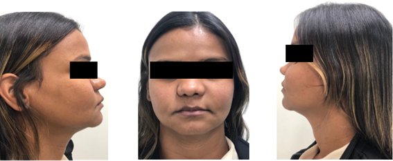
A 32-year-old female patient with no relevant systemic history was referred to the Santa Casa de Montes Claros Hospital for evaluation and removal of a bilateral palpable lesion in the maxilla. No facial asymmetry or other notable alterations were observed during the extraoral clinical examination (Figure 1). Intraoral examination revealed a mixed lesion in the maxilla, bilaterally, with suspicion of an odontogenic tumor (Figure 2).

Figure 1 Extraoral examination: lateral and frontal views where no absence of facial asymmetries was observed.
Intraoral examination revealed a pedunculated growth in the posterior maxillary region. On palpation, the swelling was firm, non-tender, and hard in consistency, with no associated bleeding or suppuration. The lesion was slow-growing, sessile, and covered by normal mucosa. Further intraoral findings included the absence of both the upper and lower anterior teeth, generalized clinical attachment loss (CAL) of 9-10 mm, and generalized probing pocket depth (PPD) of 6-7 mm. The patient was referred to the Department of Oral Pathology and Radiology for cone beam computed tomography (CBCT), which revealed extensive bone loss and bony destruction in the upper frontal region of the mandible. Additionally, the patient was referred to the Department of Stomatology and Oral Diagnostics for blood tests, including hemoglobin (Hb), bleeding time (BT), and clotting time (CT). However, no further systemic examination was possible to obtain useful complementary test results. A cone beam computed tomography scan was requested, which showed an expansive lesion in the posterior region of the right and left maxilla extending to the palatine and zygomatic processes (Figure 3).
Oral prophylaxis (ultrasonic scaling) was performed. After seven days, the patient was recalled for surgical excision of the soft tissue overgrowth. Under all aseptic conditions and precautions and under local anesthesia (lignocaine 2% and adrenaline), excision of the overgrowth was performed using a third technique, but in a conservative manner to aid in the process of repair of the surgical bed. Hemostasis was achieved using a pressure bag (gauze wrapped in local anesthesia) for 15 minutes. After achieving complete hemostasis, a dressing was performed using a periodontal pocket (Figure 4). The patient was recalled after seven days for follow-up and verification of the region where the ablative and traumatic procedure was performed.
For histopathological analysis, an incisional biopsy was performed on both sides of the maxilla. Microscopic examination using hematoxylin and eosin (H&E) staining revealed proliferation of spindle cells amid collagenized stroma, with the presence of mitoses and foci of concentric calcification, accompanied by lacerated bone trabeculae and edematous fibrous connective tissue, which were consistent with the diagnosis of odontogenic fibroma. Considering the clinical, imaging and histopathological findings, the surgical technique of enucleation followed by curettage was recommended (Figure 4). During removal, the lesion detached easily and it was necessary to remove a premolar corresponding to the area involved, which presented advanced mobility due to loss of support, followed by adequate cleaning of the surgical site.
Postoperative instructions and medications, including amoxicillin 500 mg, aceclofenac (100.0 Mg) + serration-peptidase (15.0 Mg) + paracetamol (325.0 Mg), for five days were given. The excised tissue (Figure 4) with measurement 20 x 15 mm, was preserved in a formalin-filled container and given to the Department of Oral Pathology for histopathological examination. Histopathological examination revealed a proliferation of fibroblasts with collagenous stroma and a comparatively cellular fibrous connective tissue with strands and remnants of scattered inactive odontogenic epithelium, as described in the previous paragraph. The histological diagnosis given was Bilateral Maxillary Odontogenic Fibroma.
Central ossifying fibroma is a rare condition that generally has a favorable prognosis when treated with localized excision. Calcified areas are rarely observed radiographically and typically do not affect the underlying bone. However, in our case, the presence of periodontitis contributed to noticeable bone loss. As part of the differential diagnosis, it is essential to exclude other inflammatory lesions, such as peripheral ossifying fibroma, giant cell fibroma, pyogenic granuloma, and peripheral giant cell granuloma, when diagnosing peripheral odontogenic fibroma.21 COF can easily be confused with cases of Hyperplastic Dental Follicles (HDF) and calcifying epithelial odontogenic tumors (CEOT) because they present similar histological characteristics.21–24 Therefore, the diagnosis should be made considering the radiographic, clinical, and histopathological characteristics of the lesion.22 In our case, we observed histological proliferation of fibroblasts associated with abundant odontogenic epithelial bands embedded in dense collagenous stroma with foci of dystrophic calcification, compatible with the description of WHO type of COF.23 The differentiation of HDF from COF consists of the absence of fibroblastic connective tissue. Although there may be traces of calcification, in HDF it does not resemble odontogenic material such as cementum, dysplastic dentin or osteoid.21–23
Regarding the differential diagnosis of COF, Studies9,19–24 show characterized it by the presence of amyloids, which was not observed in the histopathological evaluation of this case. The present case was diagnosed in a female patient, aged 32 years, which corroborates the incidence of the disease. The most recent literature describes its prevalence in women between the second and fourth decades of life.1,2,8 Conducted a study in Brazil with 240 cases of odontogenic tumors (OT) and only 2.1% received the diagnosis of OCT.22 Furthermore found 11 cases among 34023 did not obtain any diagnosis of OCT in the same country, among 201 cases of COF.24 Other retrospective studies have been conducted worldwide, but no cases of bilateral OCT involvement in the maxilla were found, and this may be the first reported in the literature. Central Odontogenic Fibroma is characterized by its slow and asymptomatic growth, affecting in most cases the anterior region of the maxilla with increased cortical bone.3,7,8,12–14 The present report is consistent with these characteristics, except for the location of the lesion, which, although extensive, affected the posterior region bilaterally more than the anterior region.
Radiographically, COF lesions may present similarities with cases of ameloblastoma, ameloblastic fibroma, adenomatoid odontogenic tumor, and myxoma and hyperplastic dental follicle. All of these present with uni or multilocular radiolucent lesions, well delimited with a sclerotic halo, and may present radiopaque masses suggestive of mineralized material. Sometimes, they promote displacement, tooth resorption or association with impacted teeth.2,15
In the histological profile, all analyses of neoplasms that show a fibrous component should be considered a differential diagnosis of COF. Cases of hyperplastic dental follicles, ossifying fibroma, desmoplastic fibroma, ameloblastic fibroma and myxoma are examples of differential diagnosis in H&E staining.1,12 Although bilateral involvement and non-encapsulated lesion are more present in malignant lesions, COF is a rare condition, consists of a benign odontogenic tumor that is easily detached during surgery, which occurred in the present case.
Central ossifying fibroma typically presents as a benign mass with a smooth, unencapsulated surface, usually red or pink in color, and may be sessile or pedunculated. While it can occur anywhere in the maxilla, it most commonly affects the gingiva, particularly in the molar and premolar regions, extending into deeper structures. Accurate diagnosis requires both radiographic and histological evaluation to avoid misdiagnosis. Regular follow-up is essential to monitor recurrence. This case report highlights the diagnosis, surgical treatment, and long-term follow-up of a rare central ossifying fibroma. Although benign, recurrence is uncommon. The recommended treatment is complete enucleation or curettage, and in this case, the patient was monitored for five years post-surgery through clinical and radiographic evaluations to check for any complications.
None.
The authors declared no potential conflicts of interest with respect to the research, authorship, paper, and/or publication of this article. No potential competing interest was reported by the authors.

©2024 Matos, et al. This is an open access article distributed under the terms of the, which permits unrestricted use, distribution, and build upon your work non-commercially.