Journal of
eISSN: 2373-4345


Research Article Volume 14 Issue 2
1Orthodontist, Private Practice, USA
2Clinical Associate Professor, Tufts University, School of Dental Medicine, Boston, MA Former Clinical Assistant Professor, Harvard School of Dental Medicine, USA
Correspondence: Tom C Pagonis, DDS, MS, Clinical Associate Professor, Tufts University, School of Dental Medicine, Boston, MA, USA
Received: June 05, 2023 | Published: June 23, 2023
Citation: Viazis AD, Pagonis TC. A new orthodontic diagnosis based on the cupping depth of the alveolar bone. J Dent Health Oral Disord Ther. 2023;14(2):53-61. DOI: 10.15406/jdhodt.2023.14.00595
Dentistry has made significant progress since the era when teeth were commonly extracted for dental pain, extensive dental caries, and endodontic disease. In this manuscript, we introduce the cupping depth of the alveolar bone and propose a new way to assess malalignment of teeth in orthodontics. Advancements in restorative dentistry and endodontic procedures have enabled the restoration of dental hard tissue using innovative techniques and advanced materials. However, despit e these strides in restorative dentistry, a considerable gap persists in orthodontic diagnosis and practice. Fortunately, the universal application of Edward R. Angle's arbitrary treatment goal, irrespective of individual facial and developmental morphology, is finally waning due to advancements in orthodontic technologies. FASTBRACES® Technologies introduce technical advancements and a proper knowledge base for evaluating and treating the overlooked alveolar bone architecture of malaligned teeth. The axial ("horizontal") restoration of alveolar bone is promoted, which is analogous to the sagittal ("vertical") restoration of alveolar bone in periodontal treatment planning and therapy. This approach is biologically based and consistent with a patient's natural dentition and individual genetic morphologic appearance, rather than relying on subjective or arbitrary ideals. Moreover, this approach targets a highly pathogenic periodontal microbial flora associated with malaligned teeth and linked to systemic disease. Furthermore, restoring alveolar bone is not only important for suppressing this unique pathogenic microbial flora, but it also aligns with a comprehensive strategy of successful restoration of hard tissue that is emphasized in restorative dentistry and endodontics.
Keywords: dental history, restorative dentistry, endodontics, orthodontics, alveolar bone, tooth extraction
With the advancements in technology and the accumulation of knowledge, dentistry has evolved from the common practice of tooth extraction to embrace advanced restorative dentistry and endodontic procedures.1 These advancements include the introduction of innovative dental materials such as amalgam, composite resin, and ceramics, as well as the integration of imaging and digital dentistry, which have revolutionized restorative dentistry.2
Similarly, significant progress has been made in endodontics through the development of novel biomaterials and instrumentation, offering non-surgical endodontic therapy as an effective alternative to tooth extraction for managing infected pulp spaces and endodontic diseases.3
When comparing the timelines of restorative dentistry and endodontics with orthodontics, it becomes evident that while both endodontics and restorative dentistry have made significant strides in restoring dental hard tissues, the restoration of alveolar bone in orthodontics has been comparatively neglected. This discrepancy underscores the need for further research and development to bridge this gap. The restoration of alveolar bone represents a novel and crucial aspect that requires attention in orthodontic treatment.4–9
Traditionally, orthodontics heavily relied on tooth extraction or unstable dental arch expansion, which was driven by arbitrary and unscientific treatment goals. Unfortunately, this outdated approach persists in orthodontic practice, despite notable improvements in procedures and materials. This disparity underscores the existing gap in knowledge concerning the restoration of deficient alveolar bone associated with malaligned teeth in orthodontic treatment.
By focusing on the implementation of novel techniques and methods to restore deficiencies of alveolar bone, orthodontic treatment can be more comprehensive and biologically based, by considering a patient's natural dentition and individual genetic morphologic appearance, rather than relying on subjective or arbitrary ideals. Moreover, this approach recognizes the significance of targeting the highly pathogenic periodontal microbial flora associated with malaligned teeth and its implications for systemic diseases. Addressing the restoration of alveolar bone in orthodontic treatment, not only aids in suppressing this periodontal microbial flora but also aligns with the comprehensive strategy of successful hard tissue restoration emphasized in restorative dentistry and endodontics.
Dentistry has undergone significant changes over the centuries, with many milestones marking its progress.10 The ancient Egyptians used gold wire to stabilize loose teeth and create makeshift bridges, while the ancient Romans used a mixture of iron and coral powder to fill cavities.11 During the Middle Ages, barbers and blacksmiths performed dental procedures with crude instruments and sometimes even performing dental restorations with materials like animal bones.10 The evolution of modern restorative dentistry began in the 1700s with Pierre Fauchard's publication of "Le Chirurgien Dentiste," which introduced various restorative techniques.12–14 The introduction of amalgam in the early 1800s further advanced restorative dentistry, followed by the invention of dental drills and anesthesia, which made restorative procedures less painful and more efficient.15
Endodontics, or the treatment of dental pulp, dates back to ancient civilizations.16 The formalization of endodontic treatment began in the early 20th century with the development of dental radiography and new instruments for removing infected pulp from the root canal system.17,18 The rotary instrumentation technique first developed in the 1960s allowed for faster and more efficient removal of infected pulp, followed by the introduction of apex locators, enhanced microscopy and the use of lasers in the 1990s, which made the process even more precise.16 Today, endodontic treatment is a common and routine procedure, with advanced technology making it faster, more comfortable, and more effective than ever before.16,18
The advent of anesthesia and analgesia in the mid-19th century marked a significant milestone in dentistry by allowing painless restorative and endodontic procedures. The introduction of radiographic technology in the early 20th century further advanced dentistry by providing a way to diagnose and treat dental pathosis more accurately.10,18
However, in the early 20th century, the profession faced a significant challenge with the Focal Infection Theory, which posited that dental infections could cause systemic diseases. The theory led to a dramatic increase in the number of teeth extracted for supposedly prophylactic reasons, causing many unnecessary extractions, and resulting in serious health problems. The theory had a significant impact on public health, leading to the establishment of dental hygiene programs and public health initiatives to improve dental health. Despite the widespread acceptance of the focal infection theory, there were some dentists who remained skeptical. In the 1920s and 1930s, several studies were conducted to test the theory, and the results were inconclusive. However, in the 1940s, a landmark study was conducted by Dr. Frank Billings, a prominent physician who debunked this theory, and root canal treatments became the preferred treatment for infected pulp spaces.19–22
The mid-20th century saw the introduction of fluoride, which significantly reduced the incidence of dental caries.23 In the 1960s; the first dental implants were developed, marking a significant milestone in the restoration of missing teeth. Since then, dental implant technology has continued to evolve, allowing for the restoration of teeth with near-natural function and appearance.24
Advancements in restorative dentistry have also allowed for the restoration of teeth that were previously considered beyond repair. The development of adhesive bonding techniques in the 1950s and 60s marked a significant milestone in restorative dentistry, as it allowed for the restoration of teeth with minimal removal of healthy tooth structure.25 The development of composite resins in the 1960s provided a more esthetically pleasing alternative to amalgam, and the introduction of adhesive bonding in the 1980s further improved the durability of restorations.26
The introduction of computer-aided design and manufacturing (CAD/CAM) technology in the 1980s and 90s further advanced restorative dentistry, allowing for the fabrication of highly accurate and esthetic restorations.27
Restorative dentistry has evolved significantly over time, with a focus on preserving natural teeth and restoring their function and appearance whenever possible, thanks to the use of more durable and biocompatible materials. In contrast, orthodontic treatment has traditionally focused on the movement of teeth within the existing alveolar architecture, often resulting in the extraction of teeth to create space towards an arbitrary treatment goal.28 This approach does not address and ignores the underlying deficiency in alveolar bone around malaligned teeth. In effect mainstream orthodontic treatment does not attempt to restore alveolar hard tissue as the core means of tooth alignment within the patient’s alveolar architecture.
While both fields share the common goal of improving the appearance and function of teeth, their approaches to achieving this goal have been quite different. Orthodontics also has a long history, dating back to ancient civilizations who used various methods to straighten teeth, such as the use of finger pressure and crude methods, like wedging and filing.29 Tooth extraction has also been and continues to be a common practice in orthodontics. In contrast, restorative dentistry has focused on preserving natural teeth whenever possible. While advances in orthodontic techniques and technologies have led to less reliance on tooth extraction in recent years and more on expansion techniques that maybe unstable and require permanent retention, it does not match the extent of hard tissue restoration exhibited in restorative dentistry. This is primarily due to the unscientific basis of orthodontic diagnosis and treatment planning.
For nearly a decade, the authors have challenged the scientific basis of century-plus-old orthodontic concepts which have remarkably endured to this day as the main language of diagnosis, occlusion, and treatment planning.30–40 Simply stated, orthodontic techniques and an advanced orthodontic armamentarium has rapidly evolved while clinicians and academicians seem to cling to unscientific, outdated and questionably derived concepts.
Review of late nineteenth-century orthodontics in the U.S
Late-nineteenth-century America witnessed a significant shift in the systematic descriptions of orthodontic concepts. Norman W. Kingsley’s Treatise on Oral Deformities as a Branch of Mechanical Surgery (1880) is recognized as one of the earliest works to describe orthodontics and to separate it from prosthetic dentistry.41 Kingsley made several contributions to the field, including advocating extra oral forces like occipital anchorage to align protruding teeth and treating cleft palate. However, there was little focus on occlusion or teeth malalignment since patients would undergo tooth extractions to correct dental problems.
John N. Farrar, a significant figure in nineteenth-century American orthodontics, published Irregularities of the teeth and their correction, Volume I and Volume II in 1888 and 1889, respectively.41,42 Farrar was one of the first to advocate for the importance of biologic tooth movement and noted the physiologic law governing tissues during tooth movement.42–47 However, his work was quickly replaced by Simeon Guilford’s Orthodontia, which became the standard textbook in dental schools in the U.S.47 and was unfortunately influenced with biases of the time.
Edward H. Angle is considered one of the most influential and controversial figures in late-nineteenth and early-twentieth-century American orthodontics. However, his ideal of the perfect occlusion and his preference for the facial features of the ancient Greek God Apollo have been reviewed in detail by the authors and are now recognized as flawed and outdated.48–53 Angle's approach has been criticized for perpetuating century-old facial biases and promoting unrealistic and unnecessary treatment goals for patients.30–40 Despite these criticisms and the perpetuation of outdated biases, Angle's concepts were accepted and taught at dental schools worldwide throughout the twentieth century and continue to be taught even in the present day.
In the case of Farrar and Angle, it is noteworthy that the authors did not come across a single reference in their works. While Kingsley, Guilford, Farrar, and Angle are respected figures in orthodontics, it is important to acknowledge that their contributions to the classic orthodontic literature were not grounded in rigorous scientific evidence. Instead, their writings can be seen as subjective opinions influenced by the prevailing beliefs and biases of their era. While their works undoubtedly hold historical significance, they do not align with the standards of scientific rigor that are expected in contemporary orthodontic research. Therefore, it is essential to approach their writings critically and consider the advancements in knowledge and understanding that have since emerged.
As modern orthodontics continues to evolve, it is imperative for clinicians to critically evaluate historical works and incorporate the latest scientific evidence into their practice. By doing so, they can provide the highest quality of care to patients and ensure that orthodontic treatments are based on solid scientific foundations. The authors firmly believe that it is time to appreciate the historical context of figures like Edward Angle and other clinicians from the late nineteenth century but also recognize the need to embrace biologically based approaches that are more in line with the demands and advancements of the modern world.
Since the late 19th century, orthodontics in the United States has witnessed remarkable developments and advancements. The focus shifted from using removable appliances made of gold, silver, or vulcanite with unpredictable results and lengthy treatment durations to the introduction of fixed appliances in the early 20th century. The development of the edgewise appliance system, incorporating brackets and wires, revolutionized orthodontics, allowing for precise control of tooth movement and the treatment of malocclusions. Throughout the mid-20th century, orthodontic materials continued to evolve. Stainless steel became the preferred material for orthodontic wires due to its strength and flexibility.
Advancements in adhesives led to the use of resin-based composite materials for bonding brackets to teeth, eliminating the need for individual bands.10
In the 1970s and 1980s, orthodontics saw further progress with the introduction of ceramic brackets, offering a more aesthetically pleasing alternative to traditional metal brackets. Nickel-titanium wires were also introduced, providing enhanced flexibility and shape memory.10 The late 20th century witnessed the integration of computer-aided design and manufacturing technologies (CAD/CAM) in orthodontics. This enabled the creation of custom-designed orthodontic appliances, including clear aligners, using digital impressions and three-dimensional imaging.
In recent years, esthetics has become a significant focus in orthodontics. With a multitude of clear aligner alternatives that although popular due to their esthetics, are quite limiting in their biomechanics and need more development. The integration of digital technologies has been another noteworthy advancement in orthodontics. Cone-beam computed tomography (CBCT) imaging provides detailed three-dimensional images of dentofacial structures, aiding in diagnosis and treatment planning.
Computer simulations and 3D printing have also revolutionized the fabrication of orthodontic appliances, ensuring greater precision and efficiency.6,10
In summary, orthodontics in the United States has evolved significantly since the late 19th century. The introduction of fixed appliances, advancements in materials such as stainless steel, ceramic, and nickel titanium along with the adoption of digital technologies and clear aligners have propelled the field forward. These developments have enhanced the patient experience in orthodontic care but have failed to avoid the reliance on permanent retention afterwards due to a lack of understanding of the role of alveolar bone to tooth position.
For over a century, orthodontics has largely ignored the unique soft and hard tissue anatomy and physiology of malaligned teeth, resulting in arbitrary and mythical standards for treatment planning and goals. However, modern dentistry calls for biologically based diagnostics and therapeutics that consider the morphology of the alveolar bone volume discrepancy associated with malerupted and malaligned teeth. Unfortunately, the diagnosis and treatment of alveolar bone morphology have been largely overlooked, despite the potential for localized periodontal pathologies with systemic consequences.
FASTBRACES® Technologies is a breakthrough for the scientific advancement of dentistry, addressing the restoration of alveolar bone volume discrepancies and facilitating proper positioning of teeth. Our approach to orthodontic diagnoses of malaligned teeth is based on the pretreatment clinical morphology of the alveolar bone and accompanying orientation of tooth roots. This approach utilizes alveolar bone morphology as a biologically based constant, expanding the scope of restorative dentistry to include the restoration of the alveolar bone. The restoration of alveolar bone should be considered of equal restorative value as a root canal.
Research studies show that malaligned teeth can facilitate the accumulation of bacterial plaque, contributing to gingival inflammation and the creation of a unique bacterial flora associated with greater bacterial pathogenicity in an already compromised and deficient alveolar architecture.54 Thus, the restoration of alveolar bone must be given the same attention as the restoration of other hard tissues.
Fortunately, the misguided and flawed universal application of Edward R. Angle's treatment goal to patients regardless of individual facial and developmental morphology is finally waning. With the introduction of FASTBRACES® technologies, we now have access to technical advancements and a proper knowledge base to evaluate and treat the overlooked alveolar bone architecture of malaligned teeth. The treatment approach of FASTBRACES® promotes the axial (“horizontal”) restoration of alveolar bone, analogous to the sagittal (“vertical”) restoration of alveolar bone in periodontal treatment planning and therapy. This treatment evaluation and protocol aligns with the principles of modern dentistry, which prioritize the restoration of teeth affected by caries and the preservation of root structure through conventional non-surgical or root canal treatment (Figure 1). This provides a logical and comprehensive approach of treating or restoring all hard tissues. Proper evaluation and treatment of malaligned teeth, such as with FASTBRACES®, can potentially reduce the risk of systemic health issues, including Alzheimer's disease, cardiovascular disease, diabetes, colorectal cancer, and respiratory tract infection.
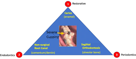
Figure 1 The three pillars of dentistry: 1- Restorative; 2-Endodontics; 3- Periodontics. The successful diagnosis and treatment of the triad of hard tissues can be seen. The yellow arrow identifies an area of hypoplasia which the authors refer to as axial or horizontal periodontics to stress that orthodontic treatment should follow in line with Restorative and Endodontics procedures, towards the restoration of alveolar bone or the third hard tissue as part of Periodontics.
A new orthodontic diagnosis based on the alveolar bone
Our orthodontic diagnoses of malaligned teeth, which were first introduced in 2017, are based on the pretreatment clinical morphology of the alveolar bone and accompanying orientation of tooth roots. This approach utilizes alveolar bone morphology as a biologically based constant, which is a logical element in the diagnostic process.37
Maxillary or mandibular alveolar hypoplasia
This represents a clinical presentation with the use of quantifying diagnostic terms of “slight”, “moderate” or “severely” crowded teeth. The level of crowding severity is no longer relevant as nearly all non-skeletal cases can be treated non-extraction. The specific loss of localized normal boney architecture and associated localized soft tissue inflammatory changes caused by malaligned roots has been termed Orthodontosis and Orthodontitis, respectively30 (Figure 1).
Maxillary or mandibular alveolar hyperplasia
While the etiology of tooth or dental spacing is multifactorial and can manifest via microdontia or the size of teeth along with physiologic habits such as thumb sucking, and tongue thrust, alveolar size is the primary factor that determines orientation of teeth. Current thought suggests that dental spacing from tongue thrust habits may be a consequence of rather than the cause of an anterior open bite.47 The clinical presentation of this diagnosis logically is spacing of teeth especially of anterior teeth with normal architecture of the alveolar bone and normal intraboney orientation of all tooth roots. Dental spacing between anterior teeth is always seen but often it is not seen with premolar teeth. One strong possibility for lack of spacing in premolar teeth is the function of the buccinator muscle with its proximity to the alveolar bone and dental arches as discussed in classic studies. Brackets are therefore often not required for teeth exhibiting normal spacing or are in proximal contact.
Addition of occlusal factors
The above referenced diagnoses would also include traditional static occlusion addendums of cross-bite, anterior open bite with specific measurements of overbite and overjet. The authors believe that recording molar relationship is not necessary particularly for a stable occlusion because the goal of orthodontic treatment should not be to change the molar relationship in pursuit of an arbitrary occlusal morphology. What’s more important is to create a functional and esthetic result by addressing an appropriate overbite/overjet of 1 to 3mm utilizing non-extraction therapy. The attainment of the overbite/overjet relation can easily be achieved in most cases via interproximal reduction (IPR) and is not within the scope of this paper. Furthermore, in cases with facial deformities in addition to the alveolar bone discrepancies of Orthodontosis described herein would of course require orthognathic surgery as the final resolution.
Assessment of alveolar bone volumetric discrepancies is an integral part of evaluating the health of periodontal tissues. Periodontal probing serves as a crucial tool in diagnosing periodontal disease. It involves the insertion of a periodontal probe into the sulcus or periodontal pocket surrounding a tooth to determine pocket depth and assess the condition of the surrounding gingival tissues.
In periodontics, the term "pocket" has evolved from its original meaning of a physical space between the tooth and the gingiva. It has become a more comprehensive and commonly used term in both periodontal diagnosis and treatment.
Similarly, the authors propose the novel diagnostic tool of alveodontal probing and the term "cup" which can be employed to describe alveolar bone hypoplasia associated with malaligned teeth (Figure 2). Like pocket depth, cupping measurement can be assessed in millimeters using a periodontal probe oriented parallel to the occlusal surface. Cupping depth offers valuable diagnostic information, including the extent of alveolar bone deficiency and the potential for future alveolar bone restoration.
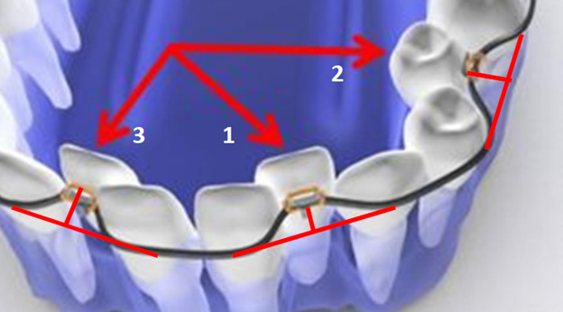
Figure 2 Graphic illustration of severely malpositioned mandibular teeth and clinical objectives of up righting (i.e., red arrows) roots to reverse Orthodontosis and eliminate Orthodontitis. Cupping diagnosis with correlating number:
The utilization of both pocketing and cupping measurements provides clinicians with a valuable diagnostic tool for assessing overall periodontal tissue health. Pocket depth and cupping depth measurements can establish diagnostic correlations with the extent of associated sagittal (‘vertical”) and axial (“horizontal”) alveolar bone loss. This novel diagnostic protocol facilitates clinicians' understanding of evaluating the axial and sagittal dimensions of alveolar bone loss and its implications for overall periodontal health.
The severity of periodontal disease is determined by the extent of bone loss and pocket depth. For instance, mild periodontitis manifests as pocket depths of 4-5 mm with minimal bone loss, while moderate periodontitis exhibits pocket depths of 6-7 mm with moderate bone loss. Severe periodontitis is characterized by pocket depths of 8 mm or more along with significant bone loss.
Based on the parameters outlined above, the authors propose diagnostic subcategories for cupping which can be described as follows (Figure 2, 3):
In effect, the greater the cupping measurement or blocking of a tooth, the greater the volumetric bone deficiency. However, unlike pocket depths in periodontal probing which generally signify greater severity of disease with increased depths, larger cupping measurements can be correlated with greater potential of alveolar bone volumetric increases to natural or normal levels.
To measure cupping depth a periodontal probe is utilized by orienting it parallel to the incisal or occlusal plane. The probe is then employed to determine the distance between the facial or buccal surface of the misaligned tooth at its most prominent facial convexity and the facial or buccal surface of the adjacent tooth at its most prominent facial convexity. This measurement provides an assessment of the depth of cupping in the maligned tooth thereby providing a quantitative assessment of alveolar bone deficiency. By incorporating these proposed diagnostic categories, clinicians can effectively assess the degree of hypoplasia based on cupping depth and the percentage of clinical crown blockage. This approach enables a comprehensive evaluation of alveolar bone discrepancies associated with hypoplasia and aids in treatment planning for optimal orthodontic tooth positioning and restoration of deficient alveolar bone.
The introduction of alveodontal probing and cupping measurements represents a significant advancement in providing a comprehensive assessment of teeth and aligning orthodontic treatment goals with the restoration of hard tissue. By incorporating quantitative analysis of alveolar bone deficiencies prior to orthodontic treatment, orthodontics moves closer to the principles of restorative dentistry and endodontics. This approach emphasizes the importance of not only achieving proper tooth alignment but also restoring the surrounding hard tissues.
Implementing alveodontal probing and cupping measurements in the dental office especially at an initial visit or as part of a dental hygiene exam can lead to increased efficiency and improved patient care.
These measurements provide valuable information for treatment planning; allowing clinicians to precisely evaluate the extent of alveolar bone deficiencies and develop appropriate treatment strategies. Furthermore, involving the dental hygienist in the active measurement and evaluation process enhances their role within the dental team and ensures a more comprehensive approach to oral health assessment and management.
By incorporating alveodontal probing and cupping measurements into routine dental practice, clinicians can enhance their ability to diagnose, plan, and monitor treatment progress. This integration of the quantitative assessment of alveolar bone facilitates, tooth alignment and the restoration of alveolar bone.
Ultimately, this promotes improved treatment outcomes and contributes to the overall success and longevity of the patient's oral health.
The associated treatment of generalized maxillary or mandibular hypoplasia consists of:
Four adult patients, seen by four different clinicians presented for orthodontic treatment with new orthodontic diagnostic terms of maxillary and mandibular hypoplasia with localized Orthodontosis and Orthodontitis (Figures 4–7),30 were successfully treated with the patented systems of FASTBRACES® Technologies. It is important to note that the universal orthodontic goal and accompanying treatment should be to successfully treat the biologically based diagnosis of the alveolar bone clinical morphology within a patient’s natural stable occlusion and morphologic appearance. Figure 4 holds particular significance as it sheds light on the complex nature of periodontal conditions, which involve multiple critical parameters. Notably, it reinforces the existing literature by demonstrating that the microbial flora surrounding tooth #25 or in an area already compromised by alveolar hypoplasia can progressively and rapidly deteriorate the alveolar bone.
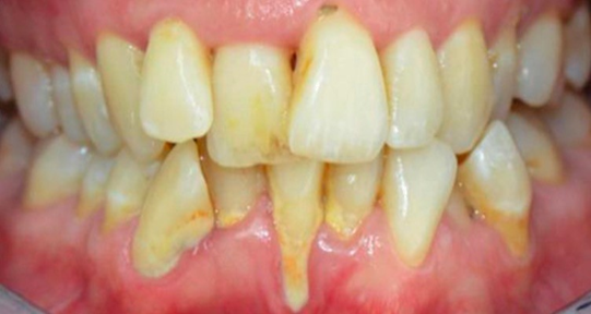
Figure 4A Before Maxillary and Mandibular Orthodontosis and Orthodontitis of anterior teeth with localized severe gingival recession. The recession of #24 may be due to hypoplasia of #23 and #25 due to the fact that alveolar bone was not enough to sustain soft tissue coverage of #24.
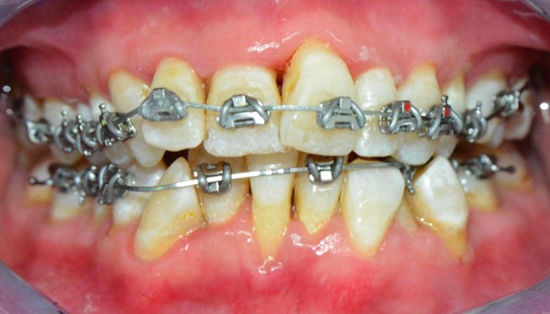
Figure 4B Brackets are placed on lingually inclined mandibular teeth to facilitate alveolar bone restoration which in turns promotes the creation healthy soft tissue and the elimination of the hypoplasia (Orthodontosis). Note that brackets are not placed on labially positioned mandibular teeth.
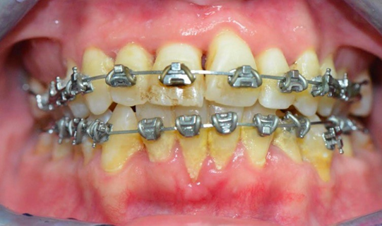
Figure 4C Bracket placement on all teeth as previously lingually inclined roots are orthoerupted (man-made eruption).
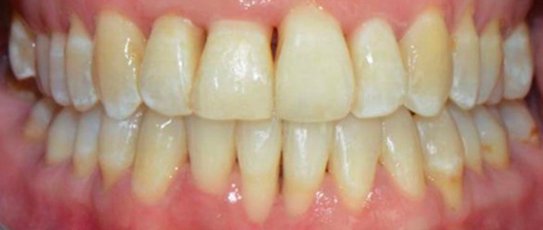
Figure 4DAfter resolution of Maxillary and Mandibular Orthodontosis and Orthodontitis with restored alveolar bone and gingival architecture.

Figure 5 A&B Pretreatment of mild cupping / alveolar bone deficiency of the maxillary right premolars (arrow) and severe cupping of the maxillary maxillary right lateral incisor (arrow).

Figure 5 C&D Post treatment of mild cupping / alveolar bone deficiency of the maxillary right premolars and severe cupping of the maxillary right lateral incisor.

Figure 6 A&B In the event of excess overjet, maxillary Interproximal reduction (IPR) is used molar to molar following space closure to reduce the overbite/overjet relation to 1 to 3mm.
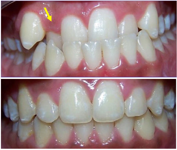
Figure 7 A&B No interproximal reduction (IPR) was needed here in the mandibular arch as the volumetric change in the alveolar bone growth in the maxillary incisor premaxilla area and especially around the maxillary right lateral, help correct the anterior crossbites/underbite. This may not be the case in more severe underbites and IPR may be needed.
However, it is important to provide additional context regarding the initial treatment approach, which involved a gingival graft to address the periodontal dehiscence. However, by forgoing traditional periodontal treatment and opting for an orthodontic alveolar bone restoration protocol with FASTBRACES® Technologies, partial coverage of the exposed tooth by alveolar bone was achieved. This suggests the convergence and potential overlap between periodontics and orthodontic alveolar bone restoration, highlighting the need for interdisciplinary collaboration. A FASTBRACES® Technologies intervention restores alveolar bone architecture, naturally aligns the teeth thereby also altering the highly pathogenic microbial flora. This is because the universal constant is the alveolar bone clinical morphology with treatment directed towards the alveolar bone deficiencies when present. These four cases are successful examples of non- extraction orthodontic treatment with the patented systems of FASTBRACES® Technologies which appropriately address the relevant deficiencies in the alveolar bone clinical morphology while maintaining a 1 to 3mm overbite /overjet and not changing the patient’s natural molar relation. The authors believe the systems of FASTBRACES® Technologies induce alveolar bone remodeling by moving the tooth roots towards their natural properly erupted positions from the onset of treatment.
The field of dentistry has made significant advancements in restoring dental hard tissue through innovative techniques and materials in restorative dentistry and endodontic procedures. However, orthodontics has lagged behind in terms of restoring dentoalveolar hard tissue, creating a notable gap in treatment approaches. Traditional orthodontic treatment planning, based on outdated facial biases from over a century ago, has often overlooked the restoration of alveolar bone deficiencies around malaligned teeth. As a result, tooth extractions have been common practice to create space for tooth alignment, which contradicts the progress made in restorative dentistry and endodontics.
To address this gap, it is crucial to implement real-world protocols for the restoration of alveolar bone in orthodontic treatment. This begins with a deep understanding of the biology of maleruption and its clinical manifestations, which then leads to the development of specific technologies and strategies to address these challenges. The authors have coined the terms "Orthodontosis" and "Orthodontitis" to describe the clinical manifestations of maligned teeth as distinct biological entities. Orthodontosis refers to the non-inflammatory deficiency or loss of volumetric alveolar bone in the horizontal dimension, typically caused by displaced tooth roots. This condition leads to excess soft tissue and chronic inflammation, known as Orthodontitis. The manuscript presents quantitative measurements, such as "cupping," to assess volumetric alveolar bone deficiencies and reviews the unique periodontal flora associated with these clinical presentations, including pathogenic gram-negative bacteria that may have systemic implications. Proper orthodontic treatment can potentially alter this pathogenic periodontal flora, providing additional protective benefits.
The authors propose that Orthodontosis and Orthodontitis result from the disruption of the natural eruption process, which causes both clinical alveolar bone volumetric deficiencies and the appearance of crowded teeth. It becomes evident that appropriate orthodontic treatment should focus on facilitating natural tooth eruption while simultaneously restoring alveolar bone to its proper architecture and volume. The authors introduced the term "Orthoeruption" to describe the orthodontic facilitation of natural tooth eruption and alveolar bone restoration. The theory of Orthoeruption involves uprighting the roots of malpositioned teeth from the beginning of orthodontic treatment, utilizing FASTBRACES® technologies, which apply light forces to stimulate alveolar bone remodeling around displaced roots and guide them into their natural position.
The manuscript extensively discusses Edward Angle's classification system and highlights the unscientific and arbitrary nature of orthodontic diagnosis and treatment planning, particularly in the early 20th century. The authors suggest that the over simplistic view of dental crowding as merely a lack of arch space, rather than considering volumetric alveolar bone deficiency caused by incomplete tooth eruption, led to this unscientific path. Angle's classifications, largely based on observations and biases of the time for the treatment of patients, do not represent a biologically based diagnostic system but rather a means of categorizing malocclusions and altering their impact. The authors question the value of some cephalometric norms or averages derived from these observations. Addressing the alveolar bone as a focused diagnosis has the potential for less reliance on cephalometric radiographs in orthodontic treatment. Requiring cephalometric radiographs before orthodontic treatment may become outdated and potentially inappropriate.
A biologically based orthodontic diagnostic approach is proposed, focusing on the pretreatment clinical morphology of the alveolar bone, and emphasizing the restoration of alveolar hard tissue. This approach considers the patient's natural dentition and individual genetic morphologic appearance, rather than subjective or arbitrary ideals. Alveolar discrepancies, such as Maxillary or Mandibular Alveolar Hypoplasia and Maxillary or Mandibular Alveolar Hyperplasia, play a crucial role in the biologically based orthodontic diagnosis, along with the functional goal of achieving 1 to 3mm of overjet/overbite.
The authors advocate for a non-extraction orthodontic treatment approach that aims to correct or improve the clinical morphology of the alveolar bone by repositioning tooth roots from the beginning of treatment, without altering the molar relation of the dentition. Recognizing the significance of restoring alveolar bone deficiencies, which directly impact the patient's natural facial morphology and overall oral health, it is crucial to emphasize the importance of a comprehensive approach.
In this regard, we propose an "axial alveolar bone restoration" approach, which focuses on restoring the axial ("horizontal") dimension of the alveolar bone in malaligned teeth. This concept aligns with the principles of periodontal treatment planning, where the restoration of alveolar bone deficiencies is a core objective. While the notion of "horizontal periodontics" may seem unconventional to periodontists, considering the potential involvement of alveolar bone deficiencies of malaligned teeth in the realm of periodontics could provide a broader perspective on interdisciplinary collaboration.
In summary, a biologically based orthodontic diagnostic approach that emphasizes the restoration of alveolar hard tissue based on the patient's natural dentition and individual genetic morphologic appearance is essential for maintaining optimal oral health and facial morphology. Incorporating innovative technologies like the FASTBRACES® system can enhance clinicians' ability to achieve non- extraction orthodontic treatment while restoring alveolar bone deficiencies. The authors propose a comprehensive approach to restore all hard tissues as the foundation of modern orthodontic treatment planning and therapy. By bridging the gap between restorative dentistry, endodontics, and orthodontics, a more holistic and effective approach can be achieved, leading to improved patient outcomes and long- term oral health.
Orthodontics has historically lagged behind restorative dentistry and endodontics in terms of advancements in the restoration of hard tissues. The reliance on tooth extraction and the neglect of alveolar bone deficiencies are major criticisms of traditional orthodontic approaches. However, with the introduction of technologies like FASTBRACES®, there is now an opportunity to bridge this gap and adopt a more comprehensive approach to orthodontic treatment.
By understanding the biologic factors contributing to tooth misalignment and utilizing quantitative analytic tools such as cupping to assess alveolar bone volumetric deficiencies, clinicians can now incorporate orthodontic diagnosis into the broader spectrum of dental diagnosis and treatment planning. This enables the restoration of hard tissues and emphasizes the importance of preserving natural teeth. The use of FASTBRACES® promotes the restoration of alveolar bone through axial alveolar bone restoration, which not only improves oral health but may also have systemic health benefits. By addressing malaligned teeth and alveolar bone deficiencies, the risk of various systemic conditions such as Alzheimer's disease, cardiovascular disease, diabetes, colorectal cancer, and respiratory tract infections can potentially be reduced.
Orthodontic diagnosis should be biologically based and centered on the clinical morphology of alveolar bone. Orthodontic treatment goals should be based on improving alveolar bone morphology by maintaining a stable occlusion irrespective of molar class.
There were no external sources of funding for this study.
The authors would like to thank the FASTBRACES® Dentist Providers that provided the cases shown herein.
The authors deny any conflict of interest for the acceptance of this concept.

©2023 Viazis, et al. This is an open access article distributed under the terms of the, which permits unrestricted use, distribution, and build upon your work non-commercially.