Journal of
eISSN: 2574-9943


Case Report Volume 9 Issue 2
Dermatology Department, Universidad Libre – Cali, Colombia
Correspondence: Johan Conquett Huertas, Dermatology Department, Universidad Libre – Cali, Colombia, Tel +573004909842
Received: April 02, 2025 | Published: April 25, 2025
Citation: Huertas JC, Saldarriaga IG, González G. Challenging the limits: an unusual case of pemphigus foliaceus in a pediatric patient. J Dermat Cosmetol. 2025;9(2):42-43. DOI: 10.15406/jdc.2025.09.00289
Pemphigus foliaceus (PF) is an autoimmune blistering dermatosis characterized by the formation of intraepidermal blisters with acantholysis and the presence of autoantibodies directed against desmoglein 1. Clinically, it manifests as small superficial blisters or vesicles that rapidly evolve into scaly and crusted erosions, typically distributed in seborrheic areas such as the scalp, face, and central trunk, usually without mucosal involvement.
We present the clinical case of a pediatric patient from southwestern Colombia diagnosed with pemphigus foliaceus, aiming to illustrate the clinical and histopathological characteristics of this uncommon disease in an age group where its presentation is rare. The importance of early diagnosis and timely management of this condition is emphasized to prevent severe complications, highlighting the diagnostic challenge posed by PF, especially in the pediatric population, where its atypical presentation can lead to initial therapeutic misjudgements and potentially fatal complications.
Keywords: pemphigus, desmoglein 1, vesiculobullous skin disease, adolescent
Pemphigus foliaceus (PF) is a chronic autoimmune blistering skin disease characterized histopathologically by intraepidermal blister formation with acantholysis and immunologically by the presence of circulating autoantibodies against the epidermis, which are responsible for its dermatological manifestations.1 This condition belongs to the group of pemphigus diseases, vesiculobullous disorders that can occur at any age, though they are more common in patients between 50 and 60 years old. However, the mean age at diagnosis may vary significantly depending on the country of origin and ethnicity. It is considered a rare disease in pediatric patients,2 with the most frequent incidence rates ranging between 0.1 and 0.5 per 100,000 people per year.3
The severity of this group of diseases lies in their progressive course, characterized by increased body catabolism with significant loss of fluids and proteins, as well as a heightened risk of secondary bacterial and viral infections, which often lead to severe sepsis and multiple organ failure. Therefore, proper diagnosis and timely management are crucial to preventing serious complications.4 Given the broad spectrum of its clinical manifestations, this pathology represents a significant diagnostic challenge in dermatology.
A 15-year-old male from a rural area of the Cauca department, Colombia, with no significant medical history, a complete vaccination schedule for his age, and no relevant epidemiological travel history. He was evaluated at the dermatology service of a high-complexity pediatric clinic in Cali, Colombia, due to a two-month history of scaly plaques on the scalp associated with pruritic vesicular lesions. The condition was initially managed in primary care as tinea capitis with topical corticosteroids and oral antifungals, without clinical improvement.
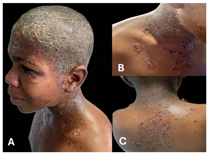
On physical examination, the patient had Fitzpatrick skin type VI. The scalp showed a widespread yellowish crust over an erythematous base. Additionally, erythematous papulovesicles, some desquamated and others with haemorrhagic crusts, were observed on the face, neck, presternal, and interscapular regions. A flaccid blister on an erythematous base was noted in the right axilla, along with isolated similar lesions on all four extremities. No mucosal involvement was observed (Figure 1).

Figure 1 Clinical photographs of the patient.
Yellowish crust on an erythematous base covering the entire thickness of the scalp. Additionally, observe some erythematous vesicles, areas of desquamation, and hemorrhagic crusts on the face (A), neck, and presternal region (B), as well as in the interscapular area (C).
Given the progression of the condition, additional laboratory tests were requested. Bloodwork results were within normal limits. KOH preparation and fungal cultures were negative. Serologic tests for HIV, HTLV I and II, and hepatitis B and C were non-reactive. Skin biopsy revealed subcorneal acantholysis with partially detached blisters, granular keratinocytes, neutrophilic exocytosis, and very few intraepidermal eosinophils. The dermis showed a perivascular lymphoplasmacytic inflammatory infiltrate with collagenization, and no microorganisms were identified in the examined samples (Figure 2). Direct immunofluorescence of perilesional skin demonstrated an intercellular reticular pattern around keratinocytes with positivity for IgG and C3, findings consistent with pemphigus foliaceus.

Figure 2 Histopathological findings.
Skin biopsy stained with hematoxylin and eosin, showing subcorneal blistering, the presence of granular keratinocytes, and marked neutrophilic exocytosis. 10X (A), 40X (B).
The patient was started on prednisone at 1 mg/kg/day for acute disease management after deworming, along with high-potency topical corticosteroid cream for skin lesions and corticosteroid lotion for scalp lesions. Follow-up at 30 days showed improvement of the blisters, with hyperpigmented macules of residual appearance at the sites of the initial lesions (Figure 3).
PF constitutes a superficial variant of pemphigus. Generally, it presents with small superficial blisters or vesicles that rapidly evolve into scaly erosions and crusted lesions, described as “bran-like”,1 which appear in a seborrheic distribution, with the scalp, face, and central trunk being most affected. Mucous membranes are typically spared, and the cutaneous lesions may be accompanied by pain or a burning sensation. Systemic symptoms are usually absent.5
For the development of the disease, both genetic and environmental factors play a crucial role in its pathogenesis. However, in patients with pemphigus, autoantibodies have been identified against a variety of epithelial cell surface antigens, intracellular components, and transmembrane elements of desmosomes,4 which could explain the range of clinical manifestations of this group of pathologies. In the specific case of PF, autoantibodies against desmoglein 1 are detected, a protein predominantly expressed in the upper layers of the epidermis. For this reason, mucous membranes generally remain unaffected due to the high levels of desmoglein 3 and the relatively low levels of desmoglein 1 expressed in them, a concept known as the “desmoglein compensation theory”.6
Two clinical variants of PF are recognized: Endemic Pemphigus Foliaceus, or Fogo Selvagem, in which an environmental trigger has been described and which occurs in rural regions of Brazil,5 and erythematous pemphigus or Senear-Usher syndrome, which describes PF manifestations localized to the malar region of the face.2
The diagnosis of PF is clinical and is confirmed by histopathological analysis with H&E staining and by direct immunofluorescence (DIF) of perilesional skin, with or without the collection of serum for indirect immunofluorescence (IIF), enzyme-linked immunosorbent assay (ELISA), or immunoblotting. Treatment is based on the prompt use of immunosuppressants such as corticosteroids (topical and systemic), steroid-sparing immunomodulators, immunoglobulins, and, more recently, anti-CD20 therapy, which has had a positive impact on the improvement and prognosis of the disease.1,2,7
In conclusion, we present a case of an infrequent and potentially severe blistering disease that generally affects adults and is very rare in the pediatric population, often being confused with other more common inflammatory or vesiculobullous disorders in this group, as occurred in the illustrated patient, who was initially managed as having a superficial mycosis due to the location of the initial lesions. Therefore, clinical suspicion and the use of appropriate diagnostic tools are essential to initiating early management and thereby reducing the morbidity and mortality associated with this condition.
None.
The authors declare there is no conflict of interest.
None.

©2025 Huertas, et al. This is an open access article distributed under the terms of the, which permits unrestricted use, distribution, and build upon your work non-commercially.