eISSN: 2574-9838


Research Article Volume 8 Issue 2
1Curso de Fisioterapia, Universidade de Araraquara - UNIARA, Brazil
2Programa de Pós-Graduação em Biotecnologia, Universidade de Araraquara - UNIARA, Brazil
Correspondence: André Capaldo Amaral, Programa de Pós-Graduação em Biotecnologia, Universidade de Araraquara - UNIARA, Brazil, Tel 55 16 99769-0095
Received: July 19, 2023 | Published: August 2, 2023
Citation: Sargentini R, Sarôa ECF, de Paula C, et al. Acute influence of myofascial self-mobilization using foam roller on muscle strength and flexibility. Int Phys Med Rehab J. 2023;8(2):155-159. DOI: 10.15406/ipmrj.2023.08.00352
Myofascial self-mobilization (MSM) techniques have been widely applied in sports, especially with the use of foam rollers. However, the effectiveness of this technique still lacks scientific consensus regarding the kinetic-functional benefits. Thirty volunteers were recruited, aged between 18 and 30 years, sedentary, and with no recent history of musculotendinous injury. The volunteers in the myofascial mobilization group (MMG/n=15) performed an MSM protocol consisting of 3 cycles of 30 seconds of mobilization. The muscle length range (MLR) assessments, established by measuring the popliteal angle, and the maximum voluntary isometric strength (MVIS) peak, by dynamometric determination, were performed before and after the protocol. The other volunteers (n=15) constituted the control group (CG) and were submitted to the same evaluation procedures, but without performing the MSM. The data obtained were submitted to descriptive qualitative analysis and the student’s t-test. The values of mean and standard deviation (M±SD) of MLR (o) before and after mobilization, respectively for the GC and MSM groups, were 157.0±8.4/158.4±9.3 and 151, 1±16.4/153.7±16.4. The peak values of MVIS (Kgf), respectively for the same groups, were 13.2±3.6/14.0±3.6 and 11.8±2.1/11.7±2.2. Given these results, it is concluded that the MSM technique with foam roller did not have an acute influence (p≥0.05) on the MLR and MVIS of the hamstring muscles.
Keywords: myofascial self-mobilization, foam roller, flexibility, muscle strength, hamstring muscles
MLR, muscle length range; MSM, myofascial self-mobilization; MMG, myofascial mobilization; CG, control group
Muscle strength and flexibility represent fundamental physical abilities, contributing to functional efficiency for maintaining a good quality of life, injury prevention and performance boost in athletes.1–3 Flexibility is determined by muscle length range (MLR) coupled with periarticular soft tissue integrity and extensibility.4 A common alternative aimed at improving flexibility, and consequently exploiting the benefits attributed to it, is muscle stretching. In recent decades, several studies have demonstrated and characterized the effectiveness of stretching programs in increasing MLR.5 Therefore, it is common to witness the inclusion of this modality in the routine of sports practice, whether recreational or competitive.6,7 In contrast, Behm and collaborators8 pointed out that despite being efficient in increasing MLR, stretching exercises can compromise performance in sports. Static stretching, one of the most used modalities in pre-exercise preparation routines, can provide acute and transient reductions in muscle strength and power levels, especially when not associated with other pre-activity modalities.9
Since static stretching can compromise sports performance, many professionals and athletes have started to adopt alternative methods to acutely increase MLR while minimizing the risks of impairment to performance.10,11 Among them, emphasis is placed on the use of the myofascial self-mobilization (MSM) technique.12 In this modality, the acute increment of MLR, and consequently of flexibility, results from the plastic changes in the myofascial structure provided by the passive stress generated in that tissue. For that, maneuvers are proposed, performed by the individual, for tissue mobilization of the fascia/muscle set, usually using auxiliary devices such as portable foam rollers, sticks or balls.13 In addition to these devices, individuals use their own body weight to apply pressure to the target musculature during the maneuver. Among the advantages attributed to MSM are its simplicity, practicality of execution, speed, and efficiency in relation to achieving its functional benefits.14,15 Although reports suggest the therapeutic effectiveness of MSM on acute MLR gain and the increasing number of practitioners, especially among competitive high-performance athletes, there is still no scientific consensus regarding the principles, parameters, and effectiveness of the technique.15 Given these facts, the present study aimed to contribute to the corpus of evidence through an investigation directed at characterizing the acute effects of the MSM technique using the foam roller on the hamstring muscles, on maximal voluntary strength and MLR.
Sample
Thirty people volunteered, aged 18 to 30 years, sedentary (minimum of 4 months prior to the research), clinically healthy and with no history of experience with the MSM technique or lower limb musculotendinous injury. To confirm their inclusion as volunteers, the participants signed a formal written consent (Informed Consent Form - ICF), according to resolution 466/2012 of the National Health Council. The research was approved by the Research Ethics Committee (registration number- 48660615.3.0000.5383). After completion of the selection stage, the volunteers were randomly divided, by drawing lots, into two groups (n=15), with one being a control group (CG) and the other assigned to the experimental procedure of myofascial mobilization (MMG).
Characterization of the sample
All the volunteers were interviewed beforehand to collect information to characterize the sample. Body mass, age, physical activity level (short version of the International Physical Activity Questionnaire - IPAq) and reports about previous experiences with the myofascial mobilization technique were obtained.
Evaluation of muscle length range (MLR)
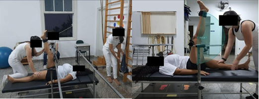
To determine the MLR, the popliteal angle measurement method was proposed. To do so, the volunteers were positioned in dorsal decubitus on a stretcher, with the hips and knees of the dominant lower limb positioned and stabilized at 90° of flexion. The contralateral lower limb was positioned with the hip and knee extended on the stretcher and stabilized by an evaluator. A strap connected to the end of an inextensible rope was attached to the distal end of the leg to be tested. The rope was attached to a pulley, fixed to a backrest, and to the opposite end was connected a receptacle (bag) containing a weight equivalent to 10% of the body mass of each volunteer. To perform the test, the receptacle was slowly released, generating, by gravitational action on the weight contained in the receptacle, a passive knee extension movement, resulting from the traction due to the redirection of the force generated by the receptacle/cord/pulley system to the lower limb under analysis. The final angular position of knee extension, established by the balance between the forces generated by the traction system and by the passive tension of the hamstring muscles, was registered in the sagittal plane by a camera (Samsung Galaxy A30s) and submitted to the determination of the angular value (popliteal angle) by the photogrammetry method. A software (Kinovea 0.9.5) was used to determine the angle by drawing two lines over the tested limb: a vertical line from the greater trochanter of the femur to the interline of the knee joint, and another line from the interline to the lateral ankle malleolus, determining the knee extension angle Figure 1.

Figure 1 Method for evaluating the muscle length range of the hamstring muscles by measuring the popliteal angle. Positioning of the volunteer and coupling of the traction device for passive knee extension (A). Establishment of the passive extension angle of the knee by photogrammetry using the Kinovea® software (B).
Evaluation of the maximum voluntary isometric strength (MVIS)
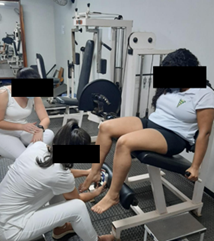
To determine the MVIS of the hamstrings, the volunteers were seated on mechanotherapy equipment (extension chair), keeping the hip and knee joints at 90° of flexion. A manual digital dynamometer (Lafayette MMT - AD instruments) was positioned between the posterior surface of the calcaneal region of the foot corresponding to the dominant limb and the support of the chair equipment intended for leg coupling Figure 2. At the signal from the evaluator, the volunteers were verbally stimulated to perform knee flexion, executing the maximum force possible, against the resistance of the device, which was locked and motionless, for 5 seconds. Three maximal voluntary isometric contractions were performed with a 1-minute interval between each contraction, and the peak MVIS value (pMVIS) of each volunteer was established by the arithmetic mean of the values established in each contraction. Before the start of the cycle of valid contractions, the volunteer performed a submaximal contraction to familiarize herself with the procedures established in this method.

Figure 2 Positioning of the volunteer for the evaluation of the peak of the voluntary isometric maximum strength of the hamstring muscles using a manual digital dynamometer (Lafayette MMT).
Myofascial self-mobilization
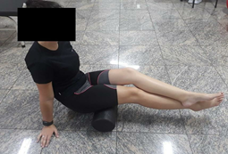
To perform the MSM technique, we used a cylindrical foam roller made of expanded polypropylene (Foam Roller Brazil), 30 cm long, 15 cm in diameter, and weighing 250 grams. The participants were initially instructed to adopt a sitting position on the ground with their lower limbs extended. Then, the roller was positioned under the posterior region of the thigh (dominant limb) and the volunteer was instructed to perform hip and knee extension of the dominant limb until the roller was the only means of contact between their lower limbs and the ground. The non-dominant limb was superimposed on the dominant and the upper limbs and trunk were actively extended, to provide stabilization and trunk elevation. From this position, the volunteers were instructed to perform, using their upper limbs as support, flexion and extension movements of trunk and hips that resulted in cyclical rolling of the posterior surface of the thigh on the roller from the region corresponding to the ischial tuberosity to the popliteal fossa. During this maneuver, the volunteers were instructed to keep the knee of the dominant limb actively extended. To standardize the mobilization execution rhythm, a rolling time of 5 seconds between the ischial tuberosity and the popliteal fossa, and vice-versa, was established, and monitored by a stopwatch (Samsung Galaxy A01). Such movements, associated with the pressure exerted by the roller on the muscles of the posterior region of the thigh, constitute the desired strategy to perform the MSM Figure 3. The MMG participants performed 3 series of MSM lasting 30s each, with 30s rest between each series. The time elapsed between performing the MLR and pMVIS tests and the MSM maneuver was 2 minutes.

Figure 3 Positioning for applying the myofascial self-mobilization technique with the foam roller on the posterior region of the thigh of the dominant limb.
Acute influence of the MSM technique
After 2 minutes, the time elapsed after finishing the application of the MSM technique, the previously described evaluation procedures of MLR and pMVIS were repeated, providing a comparative analysis of the performance of these functional parameters established in each of the volunteers before and immediately after the MSM (acute influence). The CG volunteers were submitted to the same assessment protocol, although they did not perform the MSM technique. They remained at rest for 7 minutes, a period equivalent to the time of application of self-mobilization with foam roller plus the rest intervals, drastically minimizing the influence of factors inherent to the established experimental protocol.
Statistical analysis
The values obtained from the sample characterization, MLR and MVIS measurements were submitted to qualitative descriptive analyses and the t-Student test for independent samples (comparison of pre-mean differences between the groups and post/pre differences between the two groups), considering a significance level of 5% (the difference is significant if p ≤ 0.05) to test the equality of means. Since the samples are small (15 observations for each of the two groups, GC and MGG) the validity of the normality assumption for the data necessary for using t-Student tests with small samples was checked from normal probability plots.
Sample characterization
After completing the process of selecting the volunteers, and randomly distributing them among the groups, it was possible to determine the profile of the volunteers in each group and compare them to determine the degree of homogeneity, minimizing possible interference in the experiment. The mean and standard deviation (M±SD) values for age for the CG and MMG were 21.5±2.4 and 21.9±2.3, respectively. As for body mass, the values of 64.5 ± 12.2 and 56.5 ± 6.5 were found for the CG and MMG, respectively. In both cases, the statistical analysis showed no significant difference between them.
During the experimental planning of the research, it was proposed to perform the analyses on sedentary volunteers. For this, at the selection moment, the volunteers were questioned and included only upon confirmation of their sedentary profile. To confirm such a characteristic, thus avoiding the insecurity of simply confirming the physical activity status by the volunteer's statement, it was proposed to apply the IPAq questionnaire. The results showed that, in the CG, 80% of the volunteers fit into the SEDENTARY status, and the others (20%) fit into the IRREGULARLY ACTIVE-B status. In the MMG, 93.3% of the volunteers fit into the sedentary status, and the others (6.7%) fit into the IRREGULARLY ACTIVE-B status. Within the classification scale determined by IPAq, the sedentary classification occurs when the volunteer does not perform any physical activity for at least 10 minutes continuously during the week. On the other hand, the irregularly active-B performs some type of physical activity during the week, but not enough regarding the time of execution and frequency of practice to be classified as active. Thus, we can conclude that both groups were made up of volunteers who did not have the effects of sporting practice.
Acute influence of the MLR
The results regarding the MLR in both experimental groups are illustrated in Table 1. Initially, a t-Student test for independent samples was considered to compare the pre-means in the two groups using Minitab® software (H0: mean difference = 0 versus H1: mean difference ≠ 0) and assuming the data transformed to a logarithmic scale (better normality of the data). The results do not allow rejecting H0 (p-value = 0.209 > 0.05). Similarly, the results obtained to compare the medians of the post/pre differences on the original scale for the two groups (control and MMG), also using a student’s t-test, determined the non-rejection of H0 (p-value = 0.429 > 0.05). The results obtained demonstrate, firstly, that the groups manifested homogeneity before the beginning of the experimental procedure. Furthermore, the application of the MSM technique, under the experimental conditions established in the present research, did not exert an acute influence on the MLR gain of the hamstring muscles (p ≥ 0.05). These results corroborate other studies that also showed the inexistence of the influence of this technique on MLR. Couture and collaborators16 found no changes in the MLR in a study that also investigated the effects of MSM on the hamstring muscles, with the difference in using protocols performing 4 sets of 30 seconds or 2 sets of 10 seconds in 33 active volunteers. In another study, Murray and colleagues17 evaluated the result of the application of MSM with the roller on the femoral quadriceps in 12 male athletes, performing the maneuver over 60 seconds, and also showed no significant change. However, other research has shown positive effects on MSM. Nakamura and colleagues2 concluded that performing 3 sets of 30 seconds of self-mobilization with the roller improves the MLR of the gastrocnemius muscle. They evaluated 45 sedentary volunteers in a test where discomfort tolerance determined the limit of the range of motion achieved. Similarly, the results of the research by Madoni and collaborators11 also demonstrated an increase in the flexibility of the hamstring muscles by performing 3 sets of 30 seconds of the MSM technique. It is emphasized that the time of the MSM technique applied in the present study was analogous to the time used in these studies that demonstrated increased MLR.
CG |
MMG |
||||||
Pre (°) |
Post (°) |
Pre (°) |
Post (°) |
||||
M |
157,0 |
158,4 |
151,1 |
153,7 |
|||
DP |
8,4 |
9,3 |
16,4 |
16,4 |
Table 1 Amplitude in mill volts of the Lead-1 of electrocardiography in sheep
The principal hypothesis considered to explain such a discrepancy would be the evaluation methodology of the MLR proposed in the studies. The studies in which the MSM technique provided an increase in the MLR used evaluation methods based on the volunteer's control or on the evaluator's establishment of the MLR limit, both conditions responsible for the increase in the degree of subjectivity of the methods. In the method based on the establishment of the MLR by the volunteer, the passive muscle tension limit is established by the discomfort perception. Thus, the increase in the MLR would result from a greater tolerance to the discomfort generated during the evaluation, not from the actual increase in muscle flexibility.18,19 As for methods using the evaluator as the determinant of the MLR limit, uncontrolled and disproportionate passive stresses generated in the measurements between volunteers could determine less accurate measurements. Thus, the present research elected an evaluation method in which the mechanism determining the MLR was independent of the influence of the volunteer and the evaluator, increasing the reliability of the results obtained.
Acute influence on MVIS
The results obtained on the acute effects concerning pMVIS are illustrated in Table 2. The statistical procedures used for the analysis of the pMVIS data were identical to those applied in the MLR analyses. In this case, a t-Student test for independent samples was also initially performed to compare the pre-means in the two groups, assuming the data were transformed to a logarithmic scale. The results obtained also determined the non-rejection of H0 (p-value = 0.297 > 0.05). The use of the Student's t-test to compare the means of the post/pre differences for the two groups, in the original scale, determined the non-rejection (p-value = 0.065 > 0.05) of the null hypothesis (equality of means), that is, the means of the differences are equal for CG and MMG assuming a usual significance level (5%). In addition to the verification of homogeneity between the groups before the beginning of the experimental procedures, the results demonstrate, similarly to the MLR, that the application of the MSM technique with the foam roller did not provide a significant change in the pMVIS values of the hamstring muscles (p ≥0.05), corroborating previous studies that also presented the same outcome. Madoni and coworkers,2 also using a protocol consisting of 3 sets of 30 seconds on the hamstring muscles, and Nakamura and coworkers,2 using the same protocol on the gastrocnemius muscle, reported the inexistence of the acute influence of MSM with the roller on the ability to generate maximum voluntary strength in these muscles. In the same context, but considering the strength endurance capacity using the maximum repetitions test, Simões and coworkers20 demonstrated the inability of the technique to influence the quadriceps femoris and hamstrings muscles after a MSM protocol consisting of 2 sets of 30 seconds. Taken together, the results reinforce the absence of influence of the roller MSM technique on the ability to generate maximum force in the mobilized muscle, supporting one of the advantages attributed to the technique when used before sports practice. Fonta and coworkers21 evaluated the pMVIS of the trunk extensor muscles of 25 active individuals of both sexes in a study that compared the effects of the MSM with a static stretching protocol. The results showed that the technique did not generate significant changes in strength in the pre and post-application of the technique with the foam roller.
CG |
MMG |
||||||
Pr (Kgf) |
Post (Kgf) |
Pre (Kgf) |
Post (Kgf) |
||||
M |
13,2 |
14,0 |
11,8 |
11,7 |
|||
DP |
3,6 |
3,6 |
2,1 |
2,2 |
|||
Table 2 Mean and standard deviation (M±DP) values on the peak of voluntary isometric maximum strength pre and post myofascial self-mobilization for the myofascial mobilization group (MMG). In the control group (CG) the maneuver was not performed
It is noteworthy that the objective shared by all the studies described above was to investigate the acute influence of the roller MSM technique on muscle capacities related to MLR and/or strength. If the effects of acute MLR increase without strength reduction are confirmed, as advocated by the proponents of its use, the technique could be a powerful and effective tool to contribute to sports performance improvement. On the other hand, based on existing scientific evidence to which the results presented in this research are added, it is evident that, although it does not compromise strength generation, there is no consensus on the real effectiveness of the technique to provide an improvement on the MLR, which is the main objective of its use. Given the evidence, regardless of the benefits attributed to the application of roller MSM based on the users' experience regarding the well-being provided by the maneuvers, its use should be discouraged by the inherent technical ineffectiveness. Given the current uncertainties regarding the effectiveness of the technique, new experiments should be conducted, expanding the understanding of the real influence of the maneuver execution conditions, such as different mobilization methods, the device used for mobilization, time of execution, levels of effort generated in the tissue, among others.
We conclude that the application of the foam roller MSM technique, under the experimental conditions established in this research, did not exert acute effects on the functional capacities of MLR and pMVIS of the hamstring muscles. We emphasize the importance and need for new studies capable of expanding and complementing the scientific evidence on the subject, investigating, for example, the potential influence of the parameters of technical performance to establish a consensus on the effectiveness of the technique.
None.
The authors declare that there is no conflict of interest.

©2023 Sargentini, et al. This is an open access article distributed under the terms of the, which permits unrestricted use, distribution, and build upon your work non-commercially.