International Journal of
eISSN: 2574-8084


Research Article Volume 12 Issue 3
1Department of Physics, Akdeniz University, Faculty of Science, Turkey
2Department of Radiation Oncology, Akdeniz University, Faculty of Medicine, Turkey
3Department of Radiation Oncology, University of Health Sciences, Antalya Training and Research Hospital, Turkey
Correspondence: Prof. Dr. Nina Tunçel, Department of Radiation Oncology, Faculty of Medicine, and Department of Physics, Faculty of Science, Akdeniz University, Antalya, Turkey
Received: July 21, 2025 | Published: August 4, 2025
Citation: Dağ K, Tunçel N, İnal A. QC Test for intensity-modulated X-rays using gafchromic EBT3 film and 2D ion chamber array on a single linac. Int J Radiol Radiat Ther. 2025;12(3):65-72. DOI: 10.15406/ijrrt.2025.12.00424
Accurate dosimetric verification is crucial in intensity-modulated radiotherapy (IMRT) to ensure precise dose delivery to target tissues while sparing surrounding healthy structures. This study aims to compare two widely used dosimetry tools-Gafchromic EBT3 film and the IBA MatriXX 2D ion chamber array-in terms of dose distribution accuracy and spatial resolution. A series of IMRT fields were delivered using an Elekta Synergy Platform linear accelerator, and planar dose distributions were measured using both systems. Gamma index analysis was conducted with 3%/3 mm and 2%/2 mm criteria. Results showed that both systems provided clinically acceptable agreement with planned distributions, although the Gafchromic film exhibited superior spatial resolution. These findings highlight the complementary rolls of film and ion chamber array dosimetry in modern radiotherapy QA.
Keywords: IMRT, Gafchromic EBT3 film, ion chamber array, gamma index, radiotherapy QA, planar dose verification
The clinical success of intensity-modulated radiotherapy (IMRT) is strongly dependent on the accurate delivery of highly conformal and modulated dose distributions, which are generated using inverse planning algorithms to maximize tumor control while minimizing normal tissue toxicity.1 IMRT plans typically involve steep dose gradients and multiple beam angles, making pre-treatment quality assurance (QA) an essential component of clinical implementation.2 Patient-specific QA ensures that the treatment planning system (TPS) calculations align with actual beam delivery, thereby preventing clinically significant errors.3
In modern radiotherapy QA practice, planar dosimetric verification is commonly performed using two-dimensional ion chamber arrays and radiochromic films, both of which are recommended in guidance documents such as AAPM TG-119 and TG-218.4,5 Two-dimensional (2D) ion chamber arrays, such as the IBA MatriXX, are popular for their efficiency, real-time readout, and reproducibility. However, their spatial resolution is limited by the physical size and spacing of ion chambers, which can lead to volume-averaging effects, especially in regions of steep dose gradient.6
On the other hand, Gafchromic EBT3 films offer high spatial resolution and energy independence, making them well suited for verifying complex modulated fields and small target volumes.7,8 Film dosimetry, however, is time-consuming and sensitive to handling, scanner characteristics, and calibration methodology. Despite these challenges, film remains the gold standard for high-resolution dosimetric verification, particularly in stereotactic treatments and benchmarking new technologies.9,10
Several studies have investigated the comparative performance of these two systems. For example, Devic7 outlined the advantages of Gafchromic film in capturing high-resolution dose maps in IMRT and stereotactic radiosurgery.7 Low et al.11 introduced the gamma index analysis, now the standard for spatial and dose agreement evaluation, which is employed to quantitatively compare measured and planned dose distributions. More recent works have validated the performance of ion chamber arrays under various clinical conditions but also emphasize the need for supplementary film verification in challenging geometries.12
Given these considerations, the present study aims to perform a quantitative and spatial comparison between the Gafchromic EBT3 film and the IBA MatriXX 2D ion chamber array. Using a series of IMRT plans delivered with a linear accelerator, this study evaluates both systems in terms of gamma pass rates, spatial accuracy, and operational practicality. The outcomes are intended to support decision-making in clinical QA workflows and highlight the strengths and limitations of each dosimetric tool.
Among the most common tools for two-dimensional dose verification are two-dimensional ion chamber arrays and radiochromic films. Each has unique advantages: ion chamber arrays offer rapid, easy-to-interpret results, while Gafchromic films provide high spatial resolution but require more labor-intensive handling and analysis. The goal of this study is to perform a comparative dosimetric analysis using both systems to evaluate their effectiveness and practicality in verifying IMRT dose distributions.
The study was conducted using an Elekta Synergy Platform linear accelerator equipped to deliver 6 MV photon beams. Several IMRT fields were created for quality assurance purposes using a dedicated treatment planning system (TPS), and these were delivered to a solid water-equivalent phantom under reproducible setup conditions.
Dosimetry systemsGafchromic EBT3 films were selected due to their high spatial resolution and self-developing properties. Calibration was conducted by irradiating film strips with known doses and scanning them using an Epson Expression 11000XL flatbed scanner at 300 dpi. Red channel data were analyzed using image processing software, and dose maps were generated via a calibration curve.
The MatriXX ion chamber array consists of 1020 vented parallel-plate ion chambers arranged in a 32×32 grid configuration, offering fast data acquisition. The device was placed within a MultiCube phantom and aligned using room lasers and on-board imaging. The measured dose distributions were analyzed using OmniPro I’mRT software.
Conducted tests and evaluation protocolBefore conducting the tests, the linear accelerator (linac) was calibrated according to the standard protocol, delivering 1 cGy per 1 monitor unit (MU) at a photon energy of 6 MV. For film dosimetry, EBT3-type Gafchromic films were calibrated at the same energy. Films were irradiated with doses of 20, 50, 100, 200, 400, 600, and 800 cGy, scanned using a film scanner, and imported into the analysis software. The corresponding optical density (OD) values were then correlated with the dose values delivered to generate the film calibration curve (Figure 1).
Both dosimetric systems were used to measure the planar dose distributions from the same set of tests for IMRT fields. The comparison was conducted using gamma index analysis under global criteria of 3%/3 mm and 2%/2 mm, with a dose threshold of 10%. Passing rates were calculated, and profile comparisons were performed along key dose axes. Additionally, qualitative assessments of spatial resolution and user experience were documented.
MLC leakage and transmittance test
Two 20×20 cm² fields were created, and the measurement setup along with geometric parameters, in Figure 2., 1a, 1b, 1c, and 1d illustrate the transmission through the MLCs, while 2a, 2b, 2c, and 2d show the leakage between the MLCs. A dose of 25 cGy was defined at the isocenter for both fields. First, the leaves in the X2 direction were completely closed, and the four leaves at the top of the field were fully open. Then, the leaves in the X1 direction were completely closed, and the four leaves at the top of the field were opened. The purpose of this test is to examine the transmission and interleaf leakage of the leaves in both directions.
Symmetry test in square fields
To conduct the symmetry test, square fields with dimensions of 10 × 10 cm², 5 × 5 cm², and 3 × 3 cm² were generated in the treatment planning system, as illustrated in Figure 3. The corresponding measurement setup and geometric parameters are presented in Figures 4 and 5. For each field, a dose of 100 cGy was prescribed.
MLC position accuracy test (abutting sliding fields)
In the MLC Position Accuracy Test, the collimator jaws were kept fixed while the MLC leaves were programmed to move in 2 cm intervals. Within a 20 × 20 cm² field, ten individual 2 × 20 cm² segments were created, each planned to deliver a dose of 100 cGy. The measurement setup and geometric parameters are shown in Figures 4 and 5. The segments generated in the treatment planning system (TPS) were irradiated onto both the MatriXX detector and Gafchromic EBT3 film, and a profile comparison was performed along the MLC movement direction (X‑plane), as illustrated in Figure 6.
In-plane penumbra test
In the penumbra analysis, the in‑plane test was first performed by creating six 20 × 20 cm² fields to measure the in‑plane 80%–20% penumbra. In the initial field, all leaves were closed except for the 21st leaf on the X2 side, which was fully opened. Subsequently, additional fields were generated by opening the 25th, 28th, 20th (Figure 7), 16th, and 13th leaves, respectively. Each field was irradiated with a dose of 100 cGy using the OmniPro MatriXX system and Gafchromic EBT3 film. Dose profiles were obtained along the Y‑axis, and the 80%–20% penumbra values were calculated from these profiles.
Cross-plane penumbra test
In the cross‑plane penumbra analysis, ten fields, each of which has a dimension of 1 × 20 cm², were created, moving from the X1 direction toward the X2 direction (left to right) with 1 cm spacing between them (Figure 8). The measurement setup and geometric parameters are presented in Figures 4 and 5. A dose of 100 cGy was delivered to each field. To evaluate variations in cross‑plane penumbra, the 3rd, 5th, 6th, and 8th fields were irradiated using the OmniPro MatriXX system and Gafchromic EBT3 film. Dose profiles were obtained along the horizontal direction corresponding to leaf movement, and the 80%–20% penumbra values were calculated from these profiles.
Asymmetric field test
For conducting this test, a 20×20 cm² field was created in TPS, and the MLC leaves within the field were arranged in a step-like (staircase) pattern (Figure 9.). A dose of 100 cGy was prescribed to the field.
Decreasing field size test
For this test, a five-segment field was created with dimensions of 4×20, 8×20, 12×20, 16×20, and 20×20 cm². Each segment was delivered a dose of 20 cGy. The segments expanded in the direction X2 (up to 4 cm) Figure 10., resulting in a gradient in dose intensity was obtained.
MLC tongue & groove test
The effect of the Tongue & Groove phenomenon on dose distributions within the IMRT field was investigated through this test. A 20×20 cm² field was created for the test, which was divided into two separate segments (Figure 11). Each segment received a dose of 50 cGy.
Multiple segment test in a single field
The multiple segments of sizes 2×2, 3×3, 4×4, 5×5, 8×8, and 10×10 cm² were arranged within a single field with each segment receiving a dose of 50 cGy, resulting in a total dose of 600 cGy. Figure 12 shows 5x5 cm2 subsegment in TPS arrangement. Multiple segment test irradiations for a single field test were acquired at linac and measurements applied using the OmniPro MatriXX system and Gafchromic EBT3 film.
Intensity-modulated field test
Regarding patient specific Intensity-Modulated treatment this test was organized. A total of 99 segments with varying dose weights were created within a 10×10 cm² field, and a total dose of 200 cGy was planned for this field in the TPS. Figure 13. illustrates the multi-segment field, depicting dose intensity during the film irradiation process.
Geometric set-up and evaluation method
The measurement setup and geometric parameters for both the OmniPro MatriXX and Gafchromic EBT3 film are shown in Figures 4 and 5. All tests were carried out using both the OmniPro MatriXX system and Gafchromic EBT3 film, and the differences between the two dosimetric systems were evaluated using the gamma analysis method.
Gamma pass rates for both systems were above 95% under the 3%/3 mm criteria, indicating high agreement with the planned distributions. Under the more stringent 2%/2 mm criteria, Gafchromic films maintained higher pass rates compared to the ion chamber array, particularly in regions with steep dose gradients.
Dose profiles extracted from both systems showed consistent trends, but the film data captured sharper penumbras and better resolution of high-gradient areas. The ion chamber array, while slightly less precise in spatial fidelity, allowed rapid setup, data acquisition, and interpretation, reducing the workload on medical physicists.
MLC leakage and transmission test
As shown in Table 1., the transmission and interleaf leakage tests of the right and left MLCs obtained from OmniPro MatriXX measurements were found to be consistent with each other. The average interleaf leakage and transmission values were 2.8% and 2.5%, respectively, while for the EBT3 film, these values were 2.5% and 2.3%, respectively. The comparison of left and right MLC transmission and leakage values from the OmniPro MatriXX measurements showed consistency between both sides, with average values ranging between 2.9% and 2.5%. The results indicate that the EBT3 film demonstrated slightly higher sensitivity in detecting transmission and leakage.
|
Positions on Figure 2. |
OmniPro MatriXX |
EBT3 Film |
||
|
Leaf Leakage (%) |
Leaf Transmission (%) |
Leaf Leakage (%) |
Leaf Transmission (%) |
|
|
1a/2a |
2.8 |
2.6 |
2.5 |
2.2 |
|
1b/2b |
3.2 |
2.7 |
2.7 |
2.4 |
|
1c/2c |
2.8 |
2.4 |
2.4 |
2.3 |
|
1d/2d |
2.5 |
2.3 |
2.3 |
2.1 |
Table 1 Percentage values of leakage and transmission by OmniPro MatriXX and Gafchromic EBT3 film
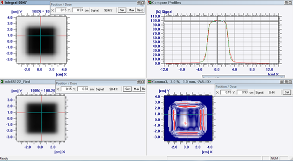
The irradiated fields on the Gafchromic EBT3 film for field sizes of 3×3 cm², 5×5 cm², and 10×10 cm² are illustrated in Figure 14. The Gamma analysis was performed using criteria of 3% dose difference and 3 mm distance-to-agreement (DTA). A notable discrepancy was observed between the results obtained from the MatriXX and the EBT3 film. As shown in Figure 15, this difference is primarily attributed to variations at the field edges. The detailed results of the Gamma Index analysis comparison between the OmniPro MatriXX and Gafchromic EBT3 film are presented in Table 2.

Figure 15 Gamma analysis comparison between Omnipro MatriXX verses Gafchromic EBT3 film for 5x5 cm2 field size.
|
Fields (cm²) |
OmniPro MatriXX–EBT3 film Gamma Index (%) |
|
10x10 |
99.05 |
|
5x5 |
99.13 |
|
3x3 |
99.47 |
Table 2 Gamma analysis results of OmniPro MatriXX verses EBT3 film
In the Gafchromic EBT3 film measurements, X-axis profiles showed minimum and maximum values of 81.4% and 85.7%, respectively (Figure 16a&16b). For OmniPro MatriXX, these values were 91% and 95%. The differences in minimum and maximum values between the two systems were 4.2% and 5%, respectively.
As shown in Table 3., the penumbra widths in the 80%–20% dose range were obtained along the Y-axis during the In-Plane Penumbra Test using both the OmniPro MatriXX and Gafchromic EBT3 film.
|
In-plane Penumbra |
OmniPro MatriXX (%) |
EBT3 film (%) |
||
|
Leaf number |
Right |
Left |
Right |
Left |
|
20 |
0.53 |
0.62 |
0.31 |
0.33 |
|
25 |
0.56 |
0.53 |
0.32 |
0.33 |
|
28 |
0.59 |
0.54 |
0.33 |
0.33 |
Table 3 Penumbra values measured along the Y-axis
As shown in Table 4., penumbra widths in the 80%–20% dose range were measured along the X-axis at various offsets during the Cross-Plane Penumbra Test using both the OmniPro MatriXX and Gafchromic EBT3 film.
|
Cross-plane Penumbra |
OmniPro MatriXX (%) |
EBT3 film (%) |
||
|
Position |
Right |
Left |
Right |
Left |
|
3 cm superior to the center |
0.86 |
1.05 |
0.48 |
0.58 |
|
Superior center |
0.63 |
0.55 |
0.52 |
0.58 |
|
Inferior center |
0.73 |
0.99 |
0.53 |
0.52 |
|
3 cm inferior to the center |
0.57 |
0.64 |
0.52 |
0.52 |
Table 4 Penumbra values measured along the X-axis
The Gamma evaluation results between the OmniPro MatriXX and Gafchromic EBT3 film showed a passing rate of 95.38%. The Gamma Index analysis presented in Figure 17. highlights the discrepancy regions in red.

Figure 17 Gamma analysis comparison between OmniPro MatriXX and Gafchromic EBT3 film for the asymmetric field test.
In this test, a profile comparison was performed along the X-axis. The comparison revealed a 92.76% agreement between the OmniPro MatriXX and Gafchromic EBT3 film. As seen in the profile comparison in Figure 18, discrepancies are present in regions 1 and 2. These differences arise from high-dose gradient areas, where the high spatial resolution of the film becomes more prominent. As the analysis moves toward the lower regions, the differences are observed to diminish.
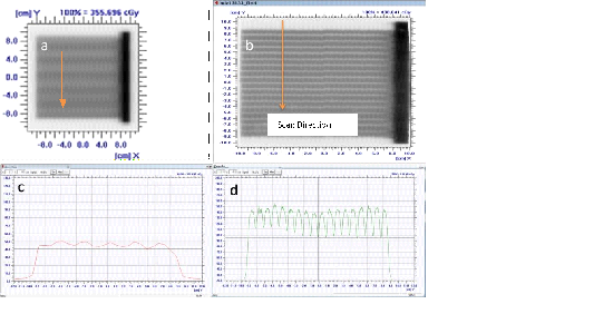
In the MLC tongue-and-groove test, the difference between the maximum and minimum points in the profile scan was found to be 18% for the OmniPro MatriXX and 34% for the Gafchromic EBT3 film (Figure 19.)

Figure 19 a) Irradiated OmniPro MatriXX image; b) Irradiated EBT3 film image; c) Profile scan along the Y-direction on OmniPro MatriXX; d) Profile scan along the Y-direction on EBT3 film.
Multiple segment test in a single field
In this test, a comparison between the OmniPro MatriXX and Gafchromic EBT3 film was performed, as shown in Figure 20. The Gamma evaluation results showed a passing rate of 99.2%.
Intensity-modulated field test
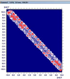
In this test, Gamma analysis was performed between the OmniPro MatriXX and Gafchromic EBT3 film, as shown in Figure 21. The Gamma Index analysis highlights the discrepancy regions in red. The Gamma evaluation yielded a passing rate of 97.10%.
Patient-specific quality assurance (QA) plays a crucial role in ensuring the accurate delivery of intensity-modulated radiotherapy (IMRT) and stereotactic radiosurgery (SRS) treatments. As treatment modalities become increasingly complex, especially in small-field, high-dose techniques like SRS and stereotactic body radiotherapy (SBRT), the demand for high-precision, spatially sensitive dosimetry systems continues to grow.
In this study, both the OmniPro MatriXX 2D ion chamber array and Gafchromic EBT3 film achieved Gamma Index pass rates above the commonly accepted clinical threshold of 95% (3%/3 mm), in line with recommendations from AAPM Task Groups TG-119 and TG-218.4,5 While these passing rates suggest that both systems are suitable for routine QA applications,13 notable differences were observed in their dosimetric resolution and sensitivity to spatial variations - especially in high- gradient regions.
Gafchromic EBT3 film showed superior performance in accurately capturing penumbra regions and leaf-edge transitions, particularly in tests such as tongue-and-groove and cross-plane penumbra evaluations. This finding is consistent with prior research by Devic et al.7 and Carrasco et al.,10 who emphasized the value of film dosimetry in scenarios where sub-millimeter resolution is required. The film’s high spatial resolution (≈ 0.1 mm) allowed it to detect small variations that were smoothed out or under-sampled by the MatriXX system, which has a detector spacing of 7.6 mm. These differences were evident in the Gamma analysis maps, where discrepancy regions (highlighted in red) often coincided with high-dose gradient zones.
Gafchromic™ EBT214 and EBT3 films are widely used in radiotherapy dosimetry. EBT3 is an improved version of EBT2, featuring a symmetric structure that eliminates scanning side dependence, which was a limitation in EBT2 due to its asymmetric design.15,16 While EBT2 shows slightly higher sensitivity, EBT3 offers better film uniformity, reduced artifacts, and greater robustness, making it more suitable for high-precision applications like SRS and IMRT QA.17,18 Additionally, EBT3’s design reduces Newton ring effects and enhances reproducibility.19
Additionally, recent studies have reaffirmed the relevance of radiochromic film in contemporary SRS QA. For instance, Wen et al.20 demonstrated that EBT3 film, when calibrated with proper multi-channel correction techniques, achieved excellent agreement with treatment planning system (TPS) calculations for SRS and SBRT plans, with Gamma pass rates exceeding 99.6% under strict 2%/2 mm criteria. Similarly, Palmer et al.21 and other recent groups22 reported that Gafchromic EBT3 and EBT‑XD film maintain high precision in high-dose, small-field applications, offering unmatched spatial accuracy for QA of complex treatment plans. These findings underscore that film remains the gold standard for verifying spatial dose accuracy in radiosurgical procedures, despite its operational complexity.
However, the use of Gafchromic film is associated with certain limitations. It requires meticulous handling, scanner calibration, and a waiting period post-irradiation to ensure dose stabilization.8,23 These requirements increase the workload and turnaround time, making it less suitable for high-throughput QA workflows.
Conversely, the OmniPro MatriXX system, while less sensitive in high-gradient regions due to its coarser detector resolution, offers significant advantages in terms of speed, ease of use, and automation. Real-time dose acquisition, integrated software analysis, and minimal setup time make it ideal for routine IMRT QA, particularly in busy clinical settings. These attributes align with the conclusions drawn by Nelms et al.3 and Nicolini et al.,24 who advocated for array-based systems as efficient tools for day-to-day QA needs.
Taken together, these results support a hybrid QA model: the OmniPro MatriXX is well-suited for routine IMRT verification due to its operational efficiency, while Gafchromic EBT3 film should be employed for high-resolution dosimetric analysis in commissioning, stereotactic plans, and investigations involving sharp dose gradients or MLC characteristics. Such a strategy leverages the strengths of both systems and complies with the best practices outlined in international QA guidelines.4,5
In conclusion, while both systems demonstrate clinical adequacy, their optimal use depends on the context of application. The MatriXX system offers practical advantages for routine QA, while film provides superior spatial accuracy essential for advanced treatment techniques. Integrating both into the QA workflow ensures comprehensive and reliable treatment verification.
Both Gafchromic EBT3 films and IBA MatriXX 2D ion chamber arrays are effective for verifying IMRT dose distributions. While the ion chamber array offers advantages in efficiency and ease of use, Gafchromic films provide enhanced spatial resolution, particularly in complex dose regions. Optimal use of these tools should be guided by clinical requirements, available resources, and case-specific QA demands.
This manuscript is based on the MSc thesis of Kadir Dağ, completed in 2018. The authors acknowledge the academic support from the Department of Physics, Akdeniz University, and technical contributions from the medical physics staff involved in QA measurements.
The authors declare no conflicts of interest related to this work.

©2025 Dağ, et al. This is an open access article distributed under the terms of the, which permits unrestricted use, distribution, and build upon your work non-commercially.