International Journal of
eISSN: 2574-8084


Case Report Volume 12 Issue 4
 Nidhi Sharma,1 Khusboo Rastogi,1 Kanika Gehlot2
1Department of Radiation Oncology, BLK Max Super speciality Hospital, India
2Department of cellular & Radiation Oncology, University of Chicago Medicine, USA
Correspondence: Deepak Mittal, Director and Head, Department of Radiation Oncology, BLK Max Super speciality Hospital, India, Tel 918-860-980-968
Received: August 27, 2025 | Published: September 9, 2025
Citation: Singh G, Mittal D, Goel V, et al. From indolent to aggressive: low-grade glioma transforming into multifocal glioblastoma with spinal metastasis. Int J Radiol Radiat Ther. 2025;12(3):92-94. DOI: 10.15406/ijrrt.2025.12.00429
Diffuse brain lesion along with leptomeningeal drop metastasis from malignant transformed high-grade glioma is rare. Here, we reported a case of a 40-year-old male who presented with malignant transformation of Grade II glioma after seven years of treatment into multifocal glioblastoma with symptomatic drop metastasis in the cervicodorsal spine. Surgical spinal decompression was done. Histopathology was suggestive of glioblastoma. Palliative radiotherapy followed by systemic therapy remains a standard approach. Patient received fractionated stereotactic radiosurgery (FSRS) to brain lesions and radiation therapy to whole spine, which he tolerated well. Relevant literature review was done regarding this rarest entity.
Keywords: Oligoastrocytoma Grade II, malignant transformation, diffuse glioma, IDH negative, 1p19q non-deleted, PNET pattern, WHO CNS classification
GBM, glioblastoma multiforme; HGG, high grade glioma; LGG, low grade glioma
Leptomeningeal metastasis from gliomas is a rare entity. Autopsy series showed that 25% of patient have spinal drop metastasis.1 Symptomatic drop metastasis is extremely rare. Here we reported a case of unusual presentation of transformed Low grade glioma to high grade glioma with multifocal intracranial recurrence with spinal drop metastasis.
A 40 years gentleman presented with headache and multiple episodes of seizures. MRI showed intraxial lesion of 5.4 × 5.4 × 6 cm. Frontal craniotomy and tumor decompression was done. Biopsy showed Oligoastrocytoma Grade II. He was given radiation (5040 cGy in 28 fractions). He was kept on follow-up. Disease-free interval was 7 years post-radiation. On follow-up, he presented with complaints of seizures after 6 years. MRI brain showed interval increase in the hyperintensity around the previous surgical cavity, suggestive of recurrence. Redo surgery was done. Biopsy showed Glioblastoma Multiforme Grade IV, IDH Negative, 1p19q non-deleted (Figure 1).
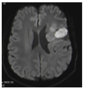
Figure 1 T1c: Thick and nodular enhancement seen along the margins of the surgical cavity.
Patient was planned for re-irradiation as DFI was 6 years. Re-irradiation was done with dose of (60 Gy/30 fractions) with Temozolomide, followed by adjuvant Temozolomide.
On follow-up, 3 months post-irradiation, patient developed weakness in right leg and decreased sensation on the back and left leg. MRI brain contrast + spine screening was done. Stable disease in insular and peri-insular region. Increase in the previous lesion in posterior body of corpus callosum. New tiny nodular lesion in left anterior frontal region. Large nodular lesion in posterior epidural space at C7–D1 levels with significant compression on the cord. Surgery was done—laminectomy and tumor excision of the C7–D1 intramedullary lesion (Figure 2a-2d).
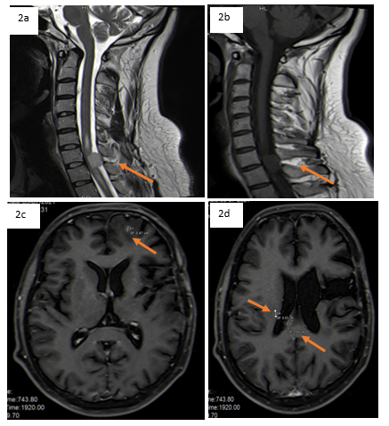
Figure 2a&2b Nodular lesion measuring 13 x 15 x 14 mm seen in the spinal canal in the posterior epidural space along the C7-D1 level.
Figure 2c&2d Rim enhancing in right half of the posterior body of the corpus callosum measuring 10.6 x 5.4 mm, body of the lateral ventricle. Tiny nodular enhancing lesion in the left anterior frontal.
Post-op HPE showed adult-type diffuse glioma, phenotype Glioblastoma with PNET pattern (WHO CNS Glioblastoma).
Multidisciplinary tumor board discussion was done. Planned for focal RT to brain lesions followed by systemic therapy (bevacizumab). Planned for whole spine radiation and focal radiation to brain lesions. Spinal irradiation (36 Gy/20# whole spine + boost to C7 lesion) up to 45 Gy, FSRT (24 Gy/3# to GTVs) to brain lesions (Figure 3&4).
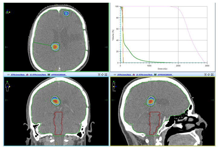
Figure 3 Colour wash showing fractionated stereotactic radiosurgery (fSRS) to brain lesions 24Gy in 3 fraction to GTV.
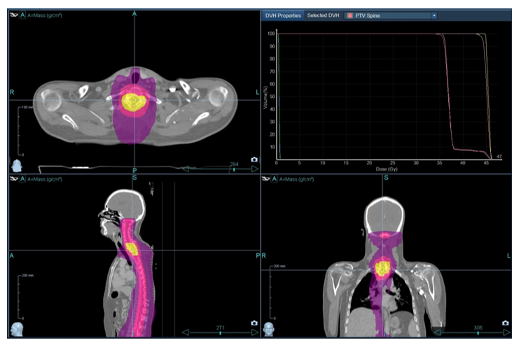
Figure 4 Colour wash showing spinal irradiation (36Gy in 20 fractions to whole spine + boost 4500 cGy to C7 lesion).
Gliomas are the most frequent type of CNS tumor. They arise from ‘glial cells’ and represent approximately 30% of all CNS tumors and 80% of all malignant CNS tumors. They can be separated into different grades (1, 2, 3, and 4) on the basis of their morphological features, histological characteristics, and molecular markers (WHO CNS 5). WHO Grade 1 and 2 are considered LGGs, while Grade 3 and 4 are HGG. WHO Grade 2 gliomas are diffusely infiltrating low-grade gliomas. They usually present in the third or fourth decade of life, and only a small proportion of patients are younger than 19 or older than 65 years. These usually arise in frontal and temporal lobes, and the most common presentation is with epileptic seizures; however, uncommonly, they may present with headache, mental changes, or lethargy. Radiologically, these are hypointense and non-enhancing on T1-weighted images and hyperintense on T2-weighted images. Since these tumors are highly infiltrative, usually the tumor extends beyond the abnormality observed on imaging. Studies have shown that MR analysis revealed an average growth rate of 5 mm/year.2
Usually, LGGs are slow-growing tumors, but the prognosis of these patients is diverse, with median survival ranging from 5 to 10 years. LGGs have the propensity to progress after resection or to undergo transformation to a higher grade (Grade 3 or 4).
Sakarunchai I et al.3 showed that the time for progression to higher grade differs for different patients, with some studies reporting 5-year malignancy-free survival rates of approximately 30 to 70%. Studies are suggesting that this malignant transformation may be due to changes in the tumor microenvironment. A study by Kaisorn L et al.4 showed that tumors with size > 3 cm and GTR were independently associated with undergoing malignant transformation. Apart from malignant transformation, these tumors may also metastasize along the CSF pathway. This leptomeningeal spread was first documented in 1931. Leptomeningeal spread is much more common in infratentorial GBM (60%) as compared to supratentorial GBM (15 to 25%), while intramedullary spinal metastases are even rarer. Ependymal invasion, fissuring of ependyma due to hydrocephalus, and repeated resections have been associated with a higher incidence of CSF dissemination.5
In our case, the tumor had undergone a malignant transformation (from Oligoastrocytoma, WHO Grade 2, to secondary Glioblastoma Multiforme, WHO Grade 4) after approximately 7 years and 4 months from the time of diagnosis. He presented with symptomatic drop metastases 3 months after completing the second course of radiation therapy for intracranial glioma. This may be attributable to a few microscopic foci of tumor cells that later presented as metachronous spinal drop metastases.
Lower thoracic, upper lumbar, and lumbosacral regions are the most common sites for spinal drop metastases in GBM, with the most common clinical presentation being radiculopathy or back pain, which may be accompanied by paraparesis or quadriparesis. Our case also had similar symptomatology.
The challenge for treating leptomeningeal metastasis is the limited treatment options. If the metastatic lesion is symptomatic, large, and causing cord compression, then surgery may be considered. But since the leptomeningeal metastases have a diffuse nature, these are usually not amenable to surgery. Thus, the mainstay of treatment for these patients is palliative radiation therapy with a total dose of 25 to 40 Gy.6
Leptomeningeal metastasis almost always results in a fatal outcome, with the average time between leptomeningeal metastasis diagnosis and death being 2 to 3 months.
To the best of my knowledge, this is the first reported case of such an unusual presentation where low-grade gliomas transformed to high-grade, then recurred with multifocal brain lesions along with spinal drop metastasis.
None.
Authors declare that there is no conflicts of interest.

©2025 Singh, et al. This is an open access article distributed under the terms of the, which permits unrestricted use, distribution, and build upon your work non-commercially.