International Journal of
eISSN: 2577-8269


Case Report Volume 4 Issue 1
1Medico Anestesiólogo del Hospital General de Sub-zona número 26, Cabo San Lucas, BCS, México
2Medico Anestesiólogo, vicepresidente mundial de anestesia intravenosa, maestro voluntario en Hospital General del Estado, Hermosillo, México
3Medico Anestesiólogo del Hospital General del Estado, Hermosillo, México
4Medico Anestesiólogo Hospital General Sub-zona número 38, San José del Cabo, México
5Medico Anestesióloga del Hospital Integral de la Mujer del Estado de Sonora, Hermosillo, México
Correspondence: Aguirre-Castro GD, Medico Anestesiólogo del Hospital General de Sub-zona número 26, Cabo San Lucas, BCS, México
Received: December 30, 2019 | Published: January 14, 2020
Citation: Aguirre-Castro GD, Rendón-Mendivil JP, Estrada-Montaño DA, et al. Erector spinae block and total intravenous anesthesia for videothoracoscopy in a patient with heart failure. Int J Fam Commun Med. 2020;4(1):1-4. DOI: 10.15406/ijfcm.2020.04.00174
68 year-old female patient, with medical history of hypertension, treated with telmisartan/hydrochlorothiazide 80/12.5/24h. Minimal effort dyspnea, orthopnea, supine hypotension, bilateral pleural effusion, pericardial effusion, heart failure with left ventricular eyection fraction (LVEF) of 20%, with very rapid evolution (2-3 months). Bilateral pleural effusions from idiopathic origin (so far). She was scheduled for diagnostic thoracoscopy, pleural effusion drainage, pericardial window, pleural and mediastinal biopsies, abrasive and chemical pleurodesis.
Anesthetic plan: Bilateral erector spinae block (ESP), total intravenous anesthesia with remifentanyl using TCI Minto model and etomidate in target manual infussion (TMI).
Conclusions: Bilateral erector spinae block showed an opioid consumption (remifentanyl) decrease to half of regular doses. Also allowing an adecuate pain management during and after surgical procedure. There were no hemodynamic variations by the time of ESP block neither with the use of etomidate in induction and maintenance of anesthesia.
Keywords: bilateral erector spinae block, regional anesthesia, intravenous anesthesia, videothorascoscopy
LVEF, left ventricular eyection fraction; ESP, erector spinae block; TMI, target manual infussion; TCI, targeted controlled infussion; Pc, plasmatic concentration; BIS, bispectral index; BP, blood pressure; HR, hearth rate; BR, breathing rate; MAP, mean arterial pressure; ESP, erector spinae plane
The use of a targeted controlled infussion (TCI) allows to maintain a low and stable plasmatic concentration (Pc) of remifentanyl during surgery, with the particularity of short onset and fast metabolism. At intubation time Pc of remifentanyl was in 2.5ng/ml with a bolus of etomidate of 13mg IV. Transanesthetic remifentanyl Pc was in a 1.75-2.5ng/ml range. Extubation was done with a remifentanyl Pc of 1.5ng/ml, the patient was awake and no pain was referred. Etomidate infussion during surgery maintained a Pc of 2ng/ml. The anesthesia depth was monetarized by bispectral index (BIS).
Ultrasound guided Erector spinae block is a relatively new analgesia technique used as a strategy in acute and chronic pain management. With multiple indications nowadays, among which, abdominothoracic surgeries stand out. The introduction of ultrasound in anesthesia has revolutionized this practice, giving great advantages like safety for the patient and the anesthesologist, allowing to watch in screen the correct spread of local anesthetic. Everyday the concept “if you can see it you can block it” gets stronger, that is cause we have neural structures as target to perform the block. The purpose in a inter fascial plane block as the ESP is to deposit the local anesthetic between anatomic spaces where the abdominal and thoracic nerves go through.1,2 Erector spinae plane block is an interfascial plane block in which local anesthetic is injected in a frontal plane of the erector spinae muscle. The anatomical references are transverse process, trapezius and rhomboid muscle.1–9 The block is performed in the thoracoabdominal region. In these case it was performed in T5 with the purpose of achieving a craniocaudal distribution about 2 spaces up and 5 under T5.3,4 The total volume of local anesthetic is about 15-20ml and it can be done as unique puncture of one side, both sides or place a catheter for continuous analgesia. The patients position can be sitting, lateral decubitus or prone. The desired level of injection is identified by counting down from the first rib (with the transducer). The ultrasound screen should be positioned in front of the operators sight, using a linear transducer placed paramedian sagittal orientation, 2 cm away from the midline; the needle insertion must be “in plane” from cephalal-to-caudal direction until the tip contacts the transverse process. To confirm proper injection plane, inject 1-3ml of local anesthetic and a spread of the erector spinae muscle, superficial to the transverse process must be seen (hydrodisection).3 The block has to be completed with 15-20ml of local anesthetic.
When a thoracic surgery is performed, the respiratory physiology in a healthy patient is going to be altered, even more in a sick patient with a pulmonary pathology. The lateral decubitus position of the patient, all anesthetics drugs, the surgery and the fact of one lung collapse lead to a ventilation/perfusion V/Q mistmach that lead to hypoxemia, functional residual capacity (FRC) decrease and diffuse interstitial edema.4 The FRC decrease worsens in post-surgical period due to pain and atelectasis. It has been demostrated that the decrease in lung volumes are about 50% in the first 24hrs and staying that way about 1-2 weeks.
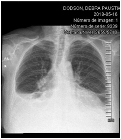
68 year-old female, born in USA, 143 pounds of weight and a 5 ft. 7 in. height; medical history of arterial hypertension, 2 years of diagnosis in pharmacological treatment with telmisartan 80mg/hydrochlorothiazide 12.5mg orally every 24hr and depression, not following the treatment. The patient arrived emergency room presenting shortness of breath with progressive minimal efforts dyspnea, orthopnea and supine hypotension. Chest x-ray: 40 % bilateral pleural effusion, mediastinal adenopathies and veiled hemidiaphragm (Figure 1). Chest tomography: cardiomegaly with global cavities increase, paratracheal adenopathies, bilateral passive atelectasis and bilateral pleural effusion (Figure 2,3).

Figure 1 Echocardiogram: dilated cardiomyophaty, left ventricular ejection fraction (LVEF) 20%, global hypokinesia, pseudonormal filling pattern, mitral moderated insufficiency, pulmonary artery systolic pressure of 57mmHg (Figure 2, 3).
Surgical plan
Diagnostic thoracoscopy, drainage of pleural effusion, pericardial window, pleural and mediastinal biopsies, abrasive and chemical pleurodesis. It is important to mention that 4hrs before surgery 300mg of hydrocortisone IV were administered to minimize adrenocortical depression incidence that etomidate infussion may cause.
Anesthetic management
Vital signs entering operating room: Blood pressure (BP): 149/91 mm/Hg, hearth rate (HR): 92 per minute, breathing rate (BR): 36 per minute, oxygen saturation (O2sat): 91 %. Temperature (T) 36.2 ºC. The patient entered operating room maintaining sitting position, always been held by her physician. With TCI system “target controlled infussion” a low remifentanyl infussion was started, a 0.2 ng/mL plasmatic concentration (P.C.), slowly increasing during one hour, until reaching P.C. of 3.0 ng/mL. Within remifentanyl start we notice a decrease of approximately 30% of mean arterial pressure (MAP). So we decided to increase the plasmatic concentration of opioid very slowly. During this time an erector spinae plane (ESP) block was performed. With patient in sitting position due to supine hypotension presented earlier, in T5 level, using Forero et al.4 technique, taking as reference C7, palpating until reaching T5, after aseptic cleaning a linear transducer (Esatote LA523 4-13 Mhz, Maastricht) with sterile cover, slided laterally 3 cm until reaching transverse process (Figure 4). The puncture was realized with BBraun 100 mm ecogenic needle, in plane, in a chephalal-to-caudal orientation. Within the contact of the transverse process 1 ml of local anestethetic was injected, evidencing hydrodisection of fascial plane between erector spinae muscle and transverse process. 130 mg of bupivacaine O.25% were administered on each side.
The patient was placed in a 30º semi fowler position. Right eyelid ptosis and bilateral miosis was observed one hour after the remifentanyl infussion, and 15 minute after the block, probably presenting Horner’s syndrome.5,6 An etomidate infussion with volumetric pump. Calculated to maintain a Pc of 2.0 ng/mL, and reaching 40% with a Biespectral index (BIS). After that, a rocuronium 35 mg bollus was administered, five minutes later, a fiber bronchoscopic selective bronchial intubation was performed and a Robert Shaw 35 endotracheal tube was placed. During the surgery, a pulmonary collapse was needed when the right pleural biopsy was taken, and when the hilar nodes where observed. The ventilator parameters were modified within requirements, and the anesthetic depth was measured with BiS to be maintained between 40-60%. Hemodynamic stability without important fluctuations of BP or HR during the surgery was achieved, managing MAP around 60 mmHg. Adjuvants: paracetamol 650 mg IV, ketorolac 60 mg IV.10
Knowing that every anesthetic medication will change our body’s hemodynamics, and the regulation systems have a very important role in that management, we were forced to look for the best option for the anesthetic procedure. The most impressive of this patient clinical presentation was the rapid onset of synthons, which progress fastly (2-3 months referred by patient) secondary to pleural and pericardial effusion, causing low effort dyspnea and orthopnea, making a conservative approach impossible.
Erector spinae block
It worked as a opioid saver, 50% approximately. The patient was hemdinamically stable, even that she had a heart failure and the block was performed billaterally, also, it helped pain management during and post-surgery, reporting a numeric rating pain scale at 1, 3, 8, 12, 18 and 24 hours post-surgery of 0, 0, 0, 3, 3, 3 respectively. The fact that the patient had no pain at the inspiration phase, allowed an adequate pulmonary compliance, with a tidal volume 0f 400-550 mL, BR 18-25 per minute, causing the risk of atelectasis decrease. Knowing that in this type of surgeries, the pulmonary volumes are around 50% lower in the first 24 hours and stay altered between one and two weeks.
Remifentanyl
The doses were 50% lower of expected, just used for the endotracheal tube and also helped an adequate plasmatic concentration at the moment of extubation for an adequate awakening, the PC was gradually decreased, until the infussion was stopped at the recovery room.
Etomidate
It’s a rapid onset hypnotic (10 sec) with a short action time (4-5 min), and a safe cardiovascular profile (7-10). At induction, no hemodynamic depression was observed. An adequate anesthetic plane was maintained with a low dose infusion.
Eyelid ptosis
Regarding the right eyelid ptosis, that settled 15 minutes after the block, a transitory Horner’s Syndrome was diagnosed. Probably followed by the sympathetic blockade.5,8 There has been no reports of this syndrome with de ESP block, however, there are multiple reports of Horner´s Syndrome after epidural block for cesarean section, with the caractheristic of transitoriness and disappearing without any complication.
Anatomopathological findings
The pleural biopsies reported acute and chronic pleuritis, with lynfoid follicular hyperplasia. Ganglia reported mixed lynphoid hyperplasia. Also hystological stains for fungi were performed, reporting negative results. The anatomopathological diagnosis was nonspecific. The clinical evolution is excellent at this moment, being self-sufficient and walking by herself without shortness of breath (Figure 5).
None.
The author declares there is no conflict of interest.

©2020 Aguirre-Castro, et al. This is an open access article distributed under the terms of the, which permits unrestricted use, distribution, and build upon your work non-commercially.