eISSN: 2373-6372


Case Report Volume 6 Issue 6
1Department of Medicine, Gleneagles Global Health City, India
2Department of Gastroenterology, Gleneagles Global Health City, India
3Department of Pathology, Gleneagles Global Health City, India
4Department of Gastrointestinal Surgery, Gleneagles Global Health City, India
Correspondence: Mayank Jain, Consultant, Department of Gastroenterology, Gleneagles Global Health City, Chennai-100, India, Tel 8959245040
Received: March 12, 2017 | Published: May 18, 2017
Citation: Madhubashini M, Jain M, Vij M, Vaithiswaran V, Chandramohan SM et al. (2017) Recurrent Partial Small Bowel Obstruction Secondary to Segmental Eosinophilic Gastroenteritis. Gastroenterol Hepatol Open Access 6(6): 00215. DOI: 10.15406/ghoa.2017.06.00215
Eosinophilic gastroenteritis is a rare disorder. The diagnostic criteria include demonstration of eosinophilic infiltration of bowel wall, lack of evidence of extra intestinal disease and exclusion of other causes of peripheral eosinophilia. We present a rare case of small bowel eosinophilic enteritis presenting as partial small bowel obstructed segment entrenched within an intra abdominal cocoon.
Eosinophilic gastroenteritis is a rare disorder thatpresents with various gastrointestinal manifestations depending on the site and specific layer of the gastrointestinal tract involved. Stomach and proximal small bowel are involved in most cases. The diagnostic criteria include demonstration of eosinophilic infiltration of bowel wall, lack of evidence of extra intestinal disease and exclusion of other causes of peripheral eosinophilia.1,2 We present a rare case of eosinophilic enteritis presenting with recurrent partial small bowel obstruction, segment embedded within an intra-abdominal cocoon.
A 31 - year - old lady presented with recurrent periumblical pain and vomiting for one year.The pain was intermittent, occurring atleast twice a day, often after a meal, and associated with abdominal distention and vomiting. The pain was aggravated often an hour after a meal and relieved by bilious non projectile vomiting. The latter was non projectile, bilious and occurred 45 minutes following a meal. There was some relief of symptoms with passage of flatus. Symptoms worsened in the following month with increasing abdominal distension, sitophobia and weight loss of 7 kgs. She was treated for pulmonary tuberculosis in the past.
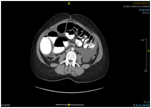
On clinical examination, patient was a febrile with normal vital parameters. The abdomen was distended; free fluid was present and gurgling was noted in left iliac fossa. Per rectal examination was normal. Her initial blood investigations including complete blood count, serum electrolytes and liver function tests were normal. X ray abdomen showed dilated small bowel loops. Chest X-ray was normal. Computed tomography scan of the abdomen showed features suggestive of sclerosing encapsulating peritonitis with subacute small bowel obstruction and a right adnexal mass (Figure 1). Ascitic fluid analysis: protein 5 g/ dL, albumin 2.3 gm/dL, total count 1200 cells/cu mm with 35 % neutrophils and 65 % eosinophils and normal adenosine deaminase levels. Upper endoscopy and colonoscopy were normal. Biopsy from the duodenum and terminal ileum did not reveal any abnormality, Mantoux test (45x25 mm) was positive. She was taken up for an emergency laparotomy.Intra-abdominal cocoon was noted andon opening the encapsulated cocoon, small intestine of approximate 30 cms length showed multiple (atleast six strictures) which was rescted (Figure 2). Histology of the cocoon capsule showedacute ulceration, fibrinoid exudate and haemorrhage with no evidence of granuloma or malignancy.The intestinal specimen showed hypertrophied muscularis propria, an edematous submucosa with mucosa showing 40 - 50 eosinophils / HPF. Subserosal region also showed dense eosinophilis (>100 eosinophils / HPF). No eosinophils were seen in the submucosa and muscularis propria. There was no evidence of granuloma, caseous necrosis, parasitic infection or vasculitis (Figure 3). Post operative period was uneventful and patient was given a short course of steroids that was tapered within 12 weeks. At present patient is asymptomatic.

Figure 1 N=57; Epidemiological distribution of the pathological fractures, traumatic fractures, and nonunion.
Eosinophilic gastroenteritis was first described by Kaijser3 Eosinophilic gastroenteritis can involve any part of gastrointestinal tract from the esophagus down to the rectum, with stomach and duodenum being the most common sites of involvement.4,5 Clinical manifestations of the disease depend on the site of eosinophil infiltration. Mucosal disease presents as protein losing enteropathy, bleeding or malabsorption, muscle layer infiltration may cause bowel wall thickening and intestinal obstruction. The subserosal form usually presents as an eosinophilic ascites. Majority of the patients give a history of seasonal allergy, atopy, food allergy, asthma and elevated Ig E levels, indicating hypersensitivity to contribute to the pathogenesis of eosinophilic enteritis.6 Presence of gastrointestinal symptoms, eosinophilic infiltration, exclusion of parasitic disease and absence of other systemic involvement with or without peripheral eosinophilia provides a clue to the diagnosis of this disorder. Dietary modification and steroids remains the mainstay of treatment. In steroid resistant cases, alternative options for management include use of mast cell stabilisers, interleukin inhibitors, mepolizumab and immunosuppression.7–10
The present case highlights a rare presentation of eosinophilic enteritis. The patient presented with intraabdominal cocoon and recurrent small bowel partial obstruction and absence of peripheral eosinophilia and atopic disorders. Though the initial ascitic fluid analysis showed a high eosinophil count, confirmation was possible on the resected segment. Both duodenal and terminal ileal biopsies were nomal. To conclude, eosinophilic enteritis is rare, it requires a high clinical index of suspicion and histological confirmation is necessary for diagnosis.
Madhubhashini M, Vaithiswaran, Mukul Vij and Chandramohan SM were involved in diagnosis and treatment. Mayank Jain and Jayanthi V were involved in management and preparation of manuscript.

©2017 Madhubashini, et al. This is an open access article distributed under the terms of the, which permits unrestricted use, distribution, and build upon your work non-commercially.